Screen Quest™ Fluo-8 Medium Removal Calcium Assay Kit *Optimized for Difficult Cell Lines*
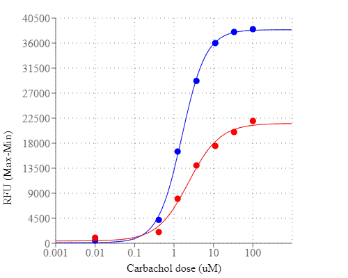
Price | |
Catalog Number | |
Unit Size | |
Quantity |
Telephone | 1-800-990-8053 |
Fax | 1-800-609-2943 |
sales@aatbio.com | |
International | See distributors |
Bulk request | Inquire |
Custom size | Inquire |
Shipping | Standard overnight for United States, inquire for international |
Correction Factor (260 nm) | 1.076 |
Correction Factor (280 nm) | 0.769 |
Extinction coefficient (cm -1 M -1) | 23430 |
Excitation (nm) | 495 |
Emission (nm) | 516 |
Quantum yield | 0.161 |
Certificate of Origin | Download PDF |
H-phrase | H303, H313, H333 |
Hazard symbol | XN |
Intended use | Research Use Only (RUO) |
R-phrase | R20, R21, R22 |
UNSPSC | 12352200 |
Overview | ![]() ![]() |
Correction Factor (260 nm) 1.076 | Correction Factor (280 nm) 0.769 | Extinction coefficient (cm -1 M -1) 23430 | Excitation (nm) 495 | Emission (nm) 516 | Quantum yield 0.161 |
Platform
Fluorescence microplate reader
Excitation | 490 nm |
Emission | 525 nm |
Cutoff | 510 nm |
Recommended plate | Black wall/clear bottom |
Instrument specification(s) | Bottom read mode/Programmable liquid handling |
Other instruments
ArrayScan, FDSS, FLIPR, FlexStation, IN Cell Analyzer, NOVOStar, ViewLuxComponents
Example protocol
AT A GLANCE
Protocol summary
- Prepare cells
- Remove the growth medium
- Add Fluo-8 NW dye working solution
- Incubate at RT for 1 hour
- Monitor fluorescence intensity at Ex/Em = 490/525 nm
Important notes
Do not add additional probenecid. It is recommended to incubate the dye working solution no longer than 2 hours.
Thaw all components to room temperature before beginning protocol.
PREPARATION OF STOCK SOLUTION
1. Fluo-8 NW stock solution:
For Cat No. 36307, add 10 µL of DMSO into Fluo-8 NW (Component A), and mix them well.
For Cat No. 36308 and 36309, add 100 µL of DMSO into Fluo-8 NW (Component A), and mix them well. Note: 10 µL of Fluo-8 NW stock solution is enough for 1 plate.
2. Assay Buffer stock solution (1X):
For Cat No. 36307 and 36308, add 9 mL of HHBS (Component C) into 10X Pluronic® F127 Plus (1 mL, Component B) and mix well.
For Cat No. 36309, add the whole bottle of 10X Pluronic® F127 Plus (10 mL, Component B) into 90 mL of HHBS buffer (not included in kit) and mix well. Note: 10 mL of 1X Assay Buffer is enough for one plate.
PREPARATION OF WORKING SOLUTION
Add 10 µL of Fluo-8 NW DMSO stock solution into 10 mL of 1X assay buffer and mix well. This working solution is stable for at least 2 hours at room temperature.
For guidelines on cell sample preparation, please visit
https://www.aatbio.com/resources/guides/cell-sample-preparation.html
SAMPLE EXPERIMENTAL PROTOCOL
- Remove the growth medium from the cell plate. Note: It is important to remove the growth medium in order to minimize background fluorescence and compound interference with serum or culture media. Note: Alternatively, grow the cells in growth medium with 0.5% - to 1% FBS to avoid medium removal step. In this case, 2X dye loading solution in HHBS buffer is needed. [We offer 2 separate no wash calcium assay kits (Cat No. 36315 and Cat No. 36316) for those who use 0.5% to 1% FBS in growth medium to avoid the medium removal step].
- Add 100 µL/well (96-well plate) or 25 µL/well (384-well plate) of Fluo-8 NW dye working solution into the cell plate.
- Incubate the dye-loading plate in a cell incubator for 30 minutes, and then incubate the plate at room temperature for another 30 minutes. Note: If the assay requires 37°C, perform the experiment immediately without further room temperature incubation. Note: If the cells can function well at room temperature for longer time, incubate the cell plate at room temperature for 1 - 2 hours (It is recommended that the incubation time be no longer than 2 hours.)
- Prepare the compound plates with HHBS or your desired buffer.
- Run the calcium flux assay by monitoring the fluorescence intensity at Ex/Em = 490/525 nm. Note: It is important to run the signal test before your experiment. Different instruments have their own intensity range. Adjust the signal test intensity to the level of 10% to 15% of the maximum intensity counts. For example, the maximum fluorescence intensity count for FLIPR-384 is 65,000, so the instrument setting should be adjusted to have its signal test intensity around 7,000 to 10,000.
Spectrum

Spectral properties
Correction Factor (260 nm) | 1.076 |
Correction Factor (280 nm) | 0.769 |
Extinction coefficient (cm -1 M -1) | 23430 |
Excitation (nm) | 495 |
Emission (nm) | 516 |
Quantum yield | 0.161 |
Images

Citations
Authors: Wu, Chung-Kuan and Wu, Chia-Lin and Lee, Tzong-Shyuan and Kou, Yu Ru and Tarng, Der-Cherng
Journal: International journal of molecular sciences (2021): 2309
Authors: Quevedo, Ana C and Lynch, Iseult and Valsami-Jones, Eugenia
Journal: Environmental Science: Nano (2021)
Authors: Ko, Hsin-Kuo and Lin, An-Hsuan and Perng, Diahn-Warng and Lee, Tzong-Shyuan and Kou, Yu Ru
Journal: Frontiers in physiology (2020): 596314
Authors: Hou, Hsin-Han and Wang, Hao-Chien and Cheng, Shih-Lung and Chen, Yen-Fu and Lu, Kai-Zen and Yu, Chong-Jen
Journal: American Journal of Physiology-Lung Cellular and Molecular Physiology (2018): L432--L442
Authors: Omotuyi, Olaposi I
Journal: Journal of Systems Biology & Proteome Research (2017)
Authors: Hirata, Yusuke and Morimoto, Yuya and Nam, Eunryel and Yoshida, Shotaro and Takeuchi, Shoji
Journal: (2017): 13--16
Authors: SHIOMI, YOSHIHIRO and YOSHIMURA, MAKOTO and KUKI, KAZUMASA and HORI, YUKO and TANAKA, TAKAO
Journal: Anticancer Research (2017): 4127--4137
Authors: Yang, Liusong and Wang, Lina and Zhu, Canjun and Wu, Junguo and Yuan, Yexian and Yu, Lulu and Xu, Yaqiong and Xu, Jingren and Wang, Tao and Liao, Zhengrui and others, undefined
Journal: Oncotarget (2017): 99470
Authors: Drzazga, Anna and Sowińska, Agata and Krzemińska, Agnieszka and Okruszek, Andrzej and Paneth, Piotr and Koziolkiewicz, Maria and Gendaszewska-Darmach, Edyta
Journal: Biochimica et Biophysica Acta (BBA)-Molecular and Cell Biology of Lipids (2017)
Authors: Wu, Jianjun and Wang, Xiangchong and Chung, Ying Ying and Koh, Cai Hong and Liu, Zhenfeng and Guo, Huicai and Yuan, Qiang and Wang, Chuan and Su, Suwen and Wei, Heming
Journal: PloS one (2017): e0168435
References
Authors: Martin VV, Beierlein M, Morgan JL, Rothe A, Gee KR.
Journal: Cell Calcium (2004): 509
Authors: Bednar B, Cunningham ME, Kiss L, Cheng G, McCauley JA, Liverton NJ, Koblan KS.
Journal: J Neurosci Methods (2004): 247
Authors: do Ceu Monteiro M, Sansonetty F, Goncalves MJ, O'Connor JE.
Journal: Cytometry (1999): 302
Authors: Cheng H, Song LS, Shirokova N, Gonzalez A, Lakatta EG, Rios E, Stern MD.
Journal: Biophys J (1999): 606
Authors: Smith GD, Keizer JE, Stern MD, Lederer WJ, Cheng H.
Journal: Biophys J (1998): 15
Authors: Su ZL, Li N, Sun YR, Yang J, Wang IM, Jiang SC.
Journal: Shi Yan Sheng Wu Xue Bao (1998): 323
Authors: Tretyn A, Kado RT, Kendrick RE.
Journal: Folia Histochem Cytobiol (1997): 41
Authors: Perez-Terzic C, Stehno-Bittel L, Clapham DE.
Journal: Cell Calcium (1997): 275
Authors: Reber BF, Schindelholz B.
Journal: Pflugers Arch (1996): 893
Authors: Greimers R, Trebak M, Moutschen M, Jacobs N, Boniver J.
Journal: Cytometry (1996): 205
Application notes
A Meta-Analysis of Common Calcium Indicators
A New Red Fluorescent & Robust Screen Quest™ Rhod-4™ Ca2+Indicator for Screening GPCR & Ca2+ Channel Targets
A New Robust No-Wash FLIPR Calcium Assay Kit for Screening GPCR and Calcium Channel Targets
A Novel NO Wash Probeniceid-Free Calcium Assay for Functional Analysis of GPCR and Calcium Channel Targets
FAQ
Are there upgraded trypan blue derivatives for cell viability testing?
Can I intracellularly measure mitochondria calcium flux and changes in mitochondria membrane potential at the same time?
Do you offer any products for measuring intracellular calcium concentration or movement by flow cytometry?
Does EDTA inactivate proteinase K?