logo
AAT Bioquest

Screen Quest™ Fluorimetric MDR Assay Kit

Effect of Cyclosporin A on the inhibition of P-gp pump in MCF-7/ADR cells. The increased concentration of Cyclosporin A resulted in an increase in fluorescence signal caused by the inhibition of P-gp pump which enhanced the intracellular accumulation of MDR indicator dye. The EC50 = 2.4 μM (measured with the kit) is similar to the value reported in the literature.
Ordering information
Price
Catalog Number
Unit Size
Quantity
Add to cart
Additional ordering information
Telephone1-800-990-8053
Fax1-800-609-2943
Emailsales@aatbio.com
InternationalSee distributors
Bulk requestInquire
Custom sizeInquire
ShippingStandard overnight for United States, inquire for international
Request quotation
Storage, safety and handling
H-phraseH303, H313, H333
Hazard symbolXN
Intended useResearch Use Only (RUO)
R-phraseR20, R21, R22
UNSPSC12352200
Related products
Screen Quest™ Membrane Potential Assay Kit *Orange Fluorescence*
Screen Quest™ Membrane Potential Assay Kit *Red Fluorescence*
Screen Quest™ 10X cell staining buffer with Phenol Red Plus™
Screen Quest™ 10X calcium assay buffer with Phenol Red Plus™
Screen Quest™ Luminometric Calcium Assay Kit
Screen Quest™ Fluo-8 Medium Removal Calcium Assay Kit *Optimized for Difficult Cell Lines*
Screen Quest™ Fluo-8 No Wash Calcium Assay Kit
Screen Quest™ Fura-2 No Wash Calcium Assay Kit
Screen Quest™ Rhod-4 No Wash Calcium Assay Kit *Medium Removal*
Screen Quest™ Rhod-4 No Wash Calcium Assay Kit
Screen Quest™ Colorimetric Chloride Channel Assay Kit
Screen Quest™ Colorimetric ELISA cAMP Assay Kit
Screen Quest™ Fluorimetric ELISA cAMP Assay Kit
Screen Quest™ TR-FRET No Wash cAMP Assay Kit
Screen Quest™ Live Cell cAMP Assay Service Pack
Screen Quest™ Fluorimetric Fatty Acid Uptake Assay Kit
Screen Quest™ Fluorimetric Glucose Uptake Assay Kit
Screen Quest™ Colorimetric Glucose Uptake Assay Kit
Screen Quest™ HEK-CNGC-Amylin 3 Receptor Cells
Screen Quest™ HEK-CNGC-Cannabinoid Receptor 1 Cells
Screen Quest™ CHO-Ga16- Chemokine (C-C) Receptor 2B Cells
Screen Quest™ HEK-CNGC-Chemokine (C-X-C motif) Receptor 4 Cells
Screen Quest™ HEK-CNGC-Dopamine Receptor 1 (DRD1) Cells
Screen Quest™ HEK-CNGC-Glucagon-like Receptor 1 (GLP1R) Cells
Screen Quest™ HEK-CNGC-Opiate Receptor-like 1 (ORL1) Cells
Screen Quest™ Human Nociceptin Receptor Ga16 coupled CHO Cells (NOP-Ga16)
Screen Quest™ CHO-Gqi Chimera Cell line
Screen Quest™ CHO-Gqo Chimera Cell line
Screen Quest™ CHO-Gqz Chimera Cell line
Screen Quest™ CHO-Gqs Chimera Cell line
Screen Quest™ CHO-Ga16 Chimera Cell line
Screen Quest™ Fluo-4 No Wash Calcium Assay Kit
Screen Quest™ No Wash Potassium Channel Assay Kit
Screen Quest™ Calbryte-520 Probenecid-Free and Wash-Free Calcium Assay Kit
Screen Quest™ Calbryte-590 Probenecid-Free and Wash-Free Calcium Assay Kit
Show More (25)

OverviewpdfSDSpdfProtocol


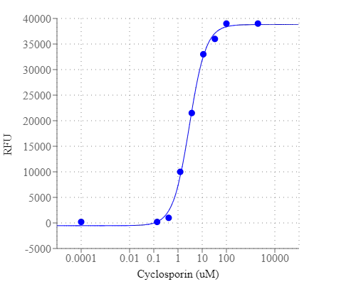
Tumor cell resistance to cytotoxic drugs is considered one of the major obstacles to successful chemotherapy. Some tumors are initially resistant and never respond to cytostatic drug treatment; others initially respond well but eventually regrow and become resistant. This phenomenon may result from genetic mutations induced by the administered antitumor agent, or may represent the selection of preexisting resistant cell populations in the malignant tumor. Multi-drug resistance (MDR) is a major factor in the failure of many forms of chemotherapy. In the past few years it has become widely accepted that the resistance to chemotherapy correlates with the overexpression of at least two ATP-dependent drug-efflux pumps. These cell membrane proteins, called P-glycoprotein (Pgp, MDR1), and multidrug-resistance-associated protein (MRP1) are members of the ABC transporter family. Our assay kit uses a fluorescent MDR indicator for assaying these two MDR pump activities. This hydrophobic fluorescent dye molecule rapidly penetrates cell membranes and becomes trapped in cells. Following a short incubation, the intracellular free dye concentration can increase significantly. In the MDR1 and/or MRP1-expressing cells this dye is extruded by the MDR transporter, thus decreasing the cellular fluorescence intensity. However, when its extrusion is blocked by an agent that interferes with the MDR1 and/or MRP1 pump-activity, its cellular fluorescence intensity increases significantly. Our MDR assay kit provides all the essential components with an optimized assay method. The assay can be performed in a convenient 96-well or 384-well microtiter-plate format and easily adapted to automation. This assay kit is ideal for high throughput screening of MDR pump inhibitors or identifying the cells that have high level of MDR pump activities.

Platform


Fluorescence microplate reader

Excitation490 nm
Emission525 nm
Cutoff515 nm
Recommended plateBlack wall/clear bottom
Instrument specification(s)Bottom read mode

Components


Example protocol


AT A GLANCE

Protocol summary

  1. Prepare cells
  2. Add MDR inhibitors or compounds
  3. Add MDR dye-loading solution (100 µL/well for 96-well plate or 25 µL/well for 384-well plate)
  4. Incubate at room temperature for 1 hour
  5. Monitor fluorescence intensity at Ex/Em = 490/525 nm with bottom read mode

Important notes
Thaw all the kit components at room temperature before use.

PREPARATION OF STOCK SOLUTION

Unless otherwise noted, all unused stock solutions should be divided into single-use aliquots and stored at -20 °C after preparation. Avoid repeated freeze-thaw cycles.

MDR sensor stock solution:
Add 20 µL (Cat. # 36340-1 plate) or 200 µL (Cat. # 36341-10 plates) of DMSO (Component B) into MDR sensor (Component A), and mix them well. Note: 20 µL of MDR sensor stock solution is enough for one plate. Un-used MDR sensor stock solution can be aliquoted and stored at < -20 oC for one month if the tubes are sealed tightly. Protect from light and avoid repeated freeze-thaw cycles and moisture.

PREPARATION OF WORKING SOLUTION

MDR dye-loading solution:
Add 20 µL of MDR sensor stock solution into 10 mL of Assay Buffer (Component C), and mix them well. Note: The MDR dye-loading solution is enough for one plate and stable for at least 2 hours at room temperature.

For guidelines on cell sample preparation, please visit
https://www.aatbio.com/resources/guides/cell-sample-preparation.html

SAMPLE EXPERIMENTAL PROTOCOL

  1. Treat cells with test compounds by adding 10 µL of 10X (96-well plate) or 5 µL of 5X (384-well plate) compounds into compound buffer (such as PBS or HHBS). For blank wells (medium without the cells), add the corresponding amount of compound buffer. Note: It is not necessary to wash cells before adding compound. However, if tested compounds are serum sensitive, growth medium and serum factors can be aspirated away before adding compounds. Add the same volume of HHBS into the wells (such as 90 µL for a 96-well plate or 20 µL for a 384-well plate) after aspiration. Alternatively, cells can be grown in serum-free media.

  2. Incubate the cell plate at room temperature or in a 37 oC, 5% CO2 incubator for at least 15 minutes or a desired period of time.

  3. Add 100 µL/well (96-well plate) or 25 µL/well (384-well plate) of MDR dye-loading solution.

  4. Incubate the dye-loading plate at room temperature for 1 hour, protected from light. Note: The appropriate incubation time depends on the individual cell type and cell concentration used. Optimize the incubation time for each experiment. (We got the optimal results with the incubation time less than 4 hours.) Note: DO NOT wash the cells after loading. Note: For non-adherent cells, it is recommended to centrifuge the cell plate at 800 rpm for 2 minutes with brake off after incubation.

  5. Monitor the fluorescence intensity at Ex/Em = 490/525 nm with bottom read mode.

Images


References


View all 77 references: Citation Explorer
Mutational Patterns Associated with the 69 Insertion Complex in Multi-drug-resistant HIV-1 Reverse Transcriptase that Confer Increased Excision Activity and High-level Resistance to Zidovudine
Authors: Cases-Gonzalez CE, Franco S, Martinez MA, Menendez-Arias L.
Journal: J Mol Biol (2007): 298
A phase I clinical and pharmacokinetic study of the multi-drug resistance protein-1 (MRP-1) inhibitor sulindac, in combination with epirubicin in patients with advanced cancer
Authors: O'Connor R, O'Leary M, Ballot J, Collins CD, Kinsella P, Mager DE, Arnold RD, O'Driscoll L, Larkin A, Kennedy S, Fennelly D, Clynes M, Crown J.
Journal: Cancer Chemother Pharmacol (2007): 79
Indomethacin overcomes doxorubicin resistance with inhibiting multi-drug resistance protein 1 (MRP1)
Authors: Matsunaga S, Asano T, Tsutsuda-Asano A, Fukunaga Y.
Journal: Cancer Chemother Pharmacol (2006): 348
Effect of therapeutic moderate hypothermia on multi-drug resistance protein 1-mediated transepithelial transport of drugs
Authors: Jin JS, Sakaeda T, Kakumoto M, Nishiguchi K, Nakamura T, Okamura N, Okumura K.
Journal: Neurol Med Chir (Tokyo) (2006): 321
Multi-Drug Resistance Conferred by Novel DNA Polymerase Mutations in Human Cytomegalovirus Isolates
Authors: Scott GM, Weinberg A, Rawlinson WD, Chou S.
Journal: Antimicrob Agents Chemother. (2006)
Synthesis of variously substituted 3-phenoxymethyl quinoxalin-2-ones and quinoxalines capable to potentiate in vitro the antiproliferative activity of anticancer drugs in multi-drug resistant cell lines
Authors: Carta A, Loriga M, Piras S, Paglietti G, La Colla P, Busonera B, Collu G, Loddo R.
Journal: Med Chem (2006): 113
Intracoronary radiation therapy for multi-drug resistant in-stent restenosis: Initial clinical experience
Authors: Price MJ, Giap H, Teirstein PS.
Journal: Catheter Cardiovasc Interv. (2006)
Editorial: the treatment of multi-drug resistant tuberculosis--a return to the pre-antibiotic era
Authors: Olle-Goig JE., undefined
Journal: Trop Med Int Health (2006): 1625
Assessment of resistance in multi drug resistant tuberculosis patients
Authors: Irfan S, Hassan Q, Hasan R.
Journal: J Pak Med Assoc (2006): 397
Orally active 1,2,4-trioxanes: synthesis and antimalarial assessment of a new series of 9-functionalized 3-(1-arylvinyl)-1,2,5-trioxaspiro[5.5]undecanes against multi-drug-resistant plasmodium yoelii nigeriensis in mice
Authors: Singh C, Malik H, Puri SK.
Journal: J Med Chem (2006): 2794