Cell Navigator™ Flow Cytometric Exosome Staining Kit
Ordering information
| Price | |
| Catalog Number | |
| Unit Size | |
| Quantity |
Additional ordering information
| Telephone | 1-800-990-8053 |
| Fax | 1-800-609-2943 |
| sales@aatbio.com | |
| International | See distributors |
| Bulk request | Inquire |
| Custom size | Inquire |
| Shipping | Standard overnight for United States, inquire for international |
Spectral properties
| Correction Factor (280 nm) | 0.35 |
| Extinction coefficient (cm -1 M -1) | 73000 |
| Excitation (nm) | 491 |
| Emission (nm) | 516 |
| Quantum yield | 0.92 |
Storage, safety and handling
| H-phrase | H303, H313, H333 |
| Hazard symbol | XN |
| Intended use | Research Use Only (RUO) |
| R-phrase | R20, R21, R22 |
| Storage | Refrigerated (2-8 °C); Minimize light exposure |
| UNSPSC | 12352200 |
Related products
| Overview |
See also: Exosomes
Correction Factor (280 nm) 0.35 | Extinction coefficient (cm -1 M -1) 73000 | Excitation (nm) 491 | Emission (nm) 516 | Quantum yield 0.92 |
Exosomes, nanoscale (30-150 nm) membrane-bound vesicles secreted by cells, are central to intercellular communication. They transfer proteins, lipids, and nucleic acids, including RNA species, between cells, playing a pivotal role in physiological and pathological processes such as immune modulation, disease progression, and genetic information exchange. This makes exosomes key targets in therapeutic and diagnostic research. However, challenges such as dye aggregation and the absence of bright, specific probes have hindered the reliable detection and analysis of exosomes. The Cell Navigator™ Flow Cytometric Exosome Staining Kit addresses these challenges by enabling the detection of CD9-positive exosomes in purified or bead-bound samples. It utilizes a fluorescent dye-conjugated CD9 marker for targeted visualization, providing a straightforward staining protocol for rapid and accurate exosome detection in diverse sample matrices, including cell culture supernatants, biological fluids, and isolated exosome preparations. To ensure effective detection and analysis, it is essential to enrich the cell culture media for exosomes before staining. This can be achieved through efficient methods such as the ReadiPrep™ Exosome Isolation Kit (Cat No. 60204) or traditional ultracentrifugation.
Platform
Flow cytometer
| Excitation | 488 nm Laser |
| Emission | 530/30 nm Filter |
| Instrument specification(s) | FITC Channel |
Example protocol
SAMPLE EXPERIMENTAL PROTOCOL
Staining of Exosomes
Isolate exosomes from cell culture medium using the ReadiPrep™ Exosome Isolation Kit (Cat# 60204) or your method of choice.
Add 5 uL of Exosome CD9 Staining Reagent into 100 uL of exosome solution.
Incubate the mix for 1 to 3 hours in the dark at room temperature.
Perform flow cytometry with FITC filter set (488 nm laser with 530/30 nm filter).
Spectrum
Open in Advanced Spectrum Viewer
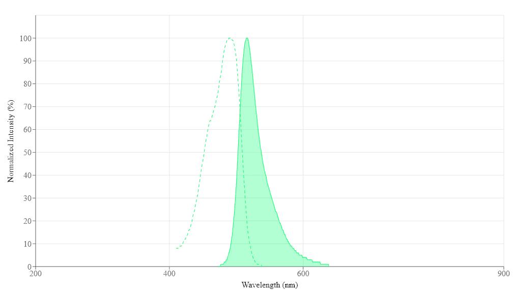
Spectral properties
| Correction Factor (280 nm) | 0.35 |
| Extinction coefficient (cm -1 M -1) | 73000 |
| Excitation (nm) | 491 |
| Emission (nm) | 516 |
| Quantum yield | 0.92 |
Images

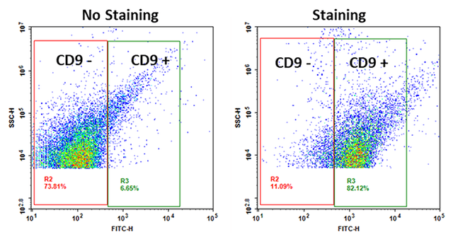
Figure 1. Exosomes from 100,000 HeLa cells (cultured for 16 hours in 10 mL serum-free medium) were isolated using ReadiPrep™ Exosome Isolation Kit (Cat# 60204) and detected using the Cell Navigator™ Flow Cytometric Exosome Staining Kit (Cat# 22426).
Application notes
A New Robust No-Wash FLIPR Calcium Assay Kit for Screening GPCR and Calcium Channel Targets
A Non-Radioactive Photometric Assay for Glucose Uptake in Insulin-Responsive 3T3-L1 Adipocytes
A Novel NO Wash Probeniceid-Free Calcium Assay for Functional Analysis of GPCR and Calcium Channel Targets
Evaluation of FLIPR Calcium Assays for Screening GPCR and Calcium Channel Targets
Restriction of Advanced Glycation End Products Improves Insulin Resistance in Human Type 2 Diabetes
FAQ
Which standard should I use with your Screen Quest Glucose Uptake Assay Kits?
What is the best way to stain cells with Membrane Marker 1-43 (MM 1-43)?
Do you have a ready-to-use cell viability assay kit?
Is there a multi-drug resistance assay kit that can detect both P-glycoprotein (P-gp, MDR1) and the multidrug resistance associated protein (MRP1)?
What assay kits measure NAD/NADH from cell samples?