Fluo-8® Calcium Indicators
In this issue
Fluorescent Calcium Indicators
Fluo-8® Calcium Indicators
Cal-520™ Calcium Indicators
Cal-590™ Calcium Indicators
Cal-630™ Calcium Indicators
Rhod-4™ Calcium Indicators
Ratiometric Calcium Indicators
FLIPR® Calcium Assays
AssayWise Letters 2015, Vol. 4(1)


Carbachol dose responses were measured in HEK-293 cells with Fluo-8® AM (Cat# 21080) and Fluo-4 AM. HEK-293 cells were seeded overnight at 40,000 cells/100 µL/well in a 96-well black wall/clear bottom Costar plate. The growth medium was removed, and the cells were incubated with 100 µL of dye-loading solution containing Fluo-8® AM or Fluo-4 AM for 1 hour at room temperature. Carbachol (25 µL/well) was added by NOVOstar to achieve the final indicated concentrations. The fluorescence signals were measured at Ex/Em = 490/525 nm. The EC50 of carbachol measured with Fluo-8® AM is about 1.2 µM.
Key Features of Fluo-8® AM:
- Faster, more readily loaded into cells than Fluo-3 AM and Fluo-4 AM. Only room temperature is required.
- Brighter, much brighter than Fluo-3 AM and Fluo-4 AM in cells.
- Convenient, almost identical spectra to those of Fluo-4 AM.


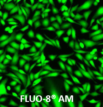
U2OS cells were seeded overnight at 40,000 cells per 100 µL per well in a Costar 96-well black wall/clear bottom plate. The growth medium was removed, and the cells were incubated with 100 µL of 4 µM Fluo-3 AM, Fluo-4 AM and Fluo-8® AM (Cat# 21080) in HHBS at 37 °C for 1 hour. The cells were washed twice with 200 µL HHBS, then imaged with a fluorescence microscope using FITC channel.
Table 1. Fluo-8® green fluorescent calcium indicators for live cell calcium imaging.
| Indicator ▲ ▼ | Ex (nm) ▲ ▼ | Em (nm) ▲ ▼ | Kd¹ ▲ ▼ | Φ² ▲ ▼ | FCa/FFree³ ▲ ▼ | Unit Size ▲ ▼ | Cat No. ▲ ▼ |
| Fluo-8®, AM | 495 | 516 | 389 nM | 0.16 | ∼200 fold | 1 mg | 21080 |
| Fluo-8®, AM | 495 | 516 | 389 nM | 0.16 | ∼200 fold | 10x50 µg | 21082 |
| Fluo-8®, AM | 495 | 516 | 389 nM | 0.16 | ∼200 fold | 20x50 µg | 21083 |
| Fluo-8®, AM | 495 | 516 | 389 nM | 0.16 | ∼200 fold | 5x50 µg | 21081 |
| Fluo-8H™, AM | 495 | 516 | 232 nM | 0.16 | ∼200 fold | 10x50 µg | 21091 |
| Fluo-8H™, AM | 495 | 516 | 232 nM | 0.16 | ∼200 fold | 1 mg | 21090 |
| Fluo-8L™, AM | 495 | 516 | 1.9 µM | 0.16 | ∼200 fold | 1 mg | 21096 |
| Fluo-8L™, AM | 495 | 516 | 1.9 µM | 0.16 | ∼200 fold | 10x50 µg | 21097 |
| Fluo-8FF™, AM | 495 | 516 | 10 µM | 0.16 | ∼200 fold | 10x50 µg | 21104 |
| Fluo-8FF™, AM | 495 | 516 | 10 µM | 0.16 | ∼200 fold | 1 mg | 21105 |