
Products
Services
Resources
Selection Guides
About
Actin Lightning™ Stains
Actin Lightning™ stains are a series of bright fluorescent probes useful for labeling F-actin in fluorescence microscopy applications, with options available for both live and fixed cell staining. This product family enables the study of actin structure and its related processes, such as cell adhesion and cell migration. Available for green, orange, and far-red channels, this product line can be paired with common nuclear counterstains and other organelle labels to enable multiplex imaging and high-plex spatial biology applications.
Cell Type | Available Colors | Example Product |
|---|---|---|
| Live Cells | Far-Red | Actin Lightning™ LC Red |
| Fixed Cells | Green, Orange, Far-Red | Actin Lightning™ iFluor® 488 Phalloidin Conjugate |
Fig. 1
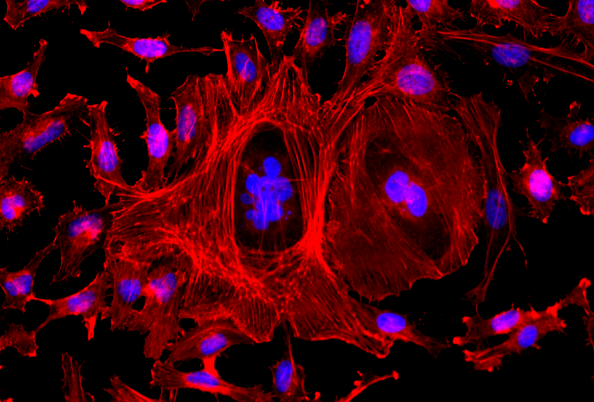
Fluorescence image of HeLa cells stained with Actin Lightning™ iFluor® 647 Phalloidin Conjugate and Nuclear Blue™ DCS1. Cells were fixed with 4% formaldehyde, permeabilized with 0.2% Triton™ X-100, then stained with Actin Lightning™ iFluor® 647 Phalloidin Conjugate (Catalog Number 22719, Red) for F-actin detection and counterstained with Nuclear Blue™ DCS1 (Catalog Number 17548, Blue). Images were acquired using Cy5 and DAPI filter sets and overlaid to show F-actin in red and nuclei in blue.
Unlike traditional phalloidin-based probes, which cannot pass through intact cell membranes, Actin Lightning™ LC Red has been designed as a cell-permeable probe that, upon entry into live cells, binds selectively to actin filaments, enabling visualization of cytoskeletal architecture in living cells without the need for fixation during staining. With an emission in the far-red Cy5 channel, Actin Lightning™ LC Red has the advantage of decreased autofluorescence, as well as being compatible with green channel multiplexing. Compared to other live cell actin stains on the market, Actin Lightning™ LC Red also demonstrates significantly brighter fluorescence under similar experimental conditions, while removing the need for any efflux pump inhibitors (such as probenecid or verapamil) to maintain intracellular signal. Finally, Actin Lightning™ LC Red is fixable, allowing staining to be retained for workflows that require cell fixation downstream.
- Cell-permeable: stains F-actin in living cells
- Strong fluorescence: significantly brighter than comparable probes like SiR-Actin
- No efflux pump inhibitor required: works without probenecid or verapamil
- Far-red (Cy5) channel: low autofluorescence, compatible with green channel multiplex
- Fixable probe: compatible with workflows requiring cell fixation
The figure below demonstrates that, under similar experimental conditions, Actin Lightning™ LC Red produces significantly brighter fluorescence staining than other live cell actin labels, such as SiR-Actin. The ability to obtain highly fluorescent images with shorter exposure times can help reduce cell phototoxicity and dye photobleaching, enabling clearer live cell imaging and more reliable analysis of dynamic actin structures.
Fig. 2
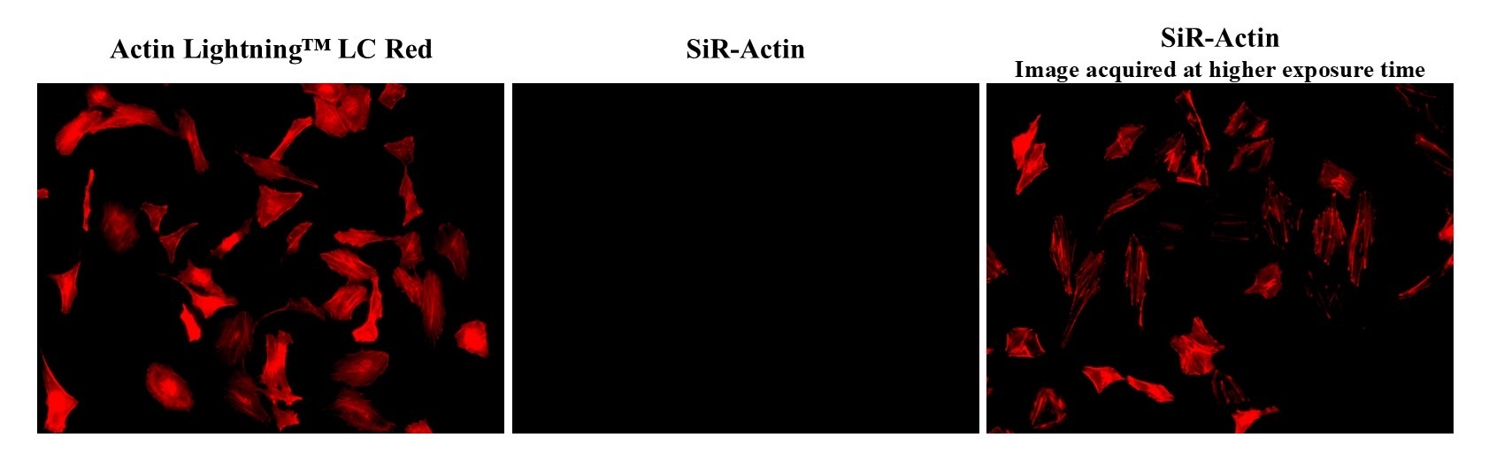
Fluorescence images of live HeLa cells stained with Actin Lightning™ LC Red (Catalog Number 22705) or SiR-Actin using fluorescence microscope with a Cy5 filter set. The image shown on far left and center was acquired at same exposure time (Exposure time = 1/3 sec) with Cy5 filter. The image on the right used much higher exposure time (Exposure time = 2 sec) with Cy5 filter. SiR-Actin does show very faint staining when image was acquired at higher exposure time.
Many live cell actin probes have poor cellular retention due to the activity of cellular efflux pumps, which causes probe leakage, leading to a loss of signal specificity and intensity while increasing background fluorescence. These probes, therefore, require the addition of efflux pump inhibitors, such as probenecid or verapamil, during experimentation. Usage of such inhibitors, however, may impact cellular function and cause cytotoxicity. As seen in the figure below, Actin Lightning™ LC Red was evaluated with and without probenecid, showing excellent cellular retention and signal quality without the usage of efflux pump inhibitors.
Fig. 3
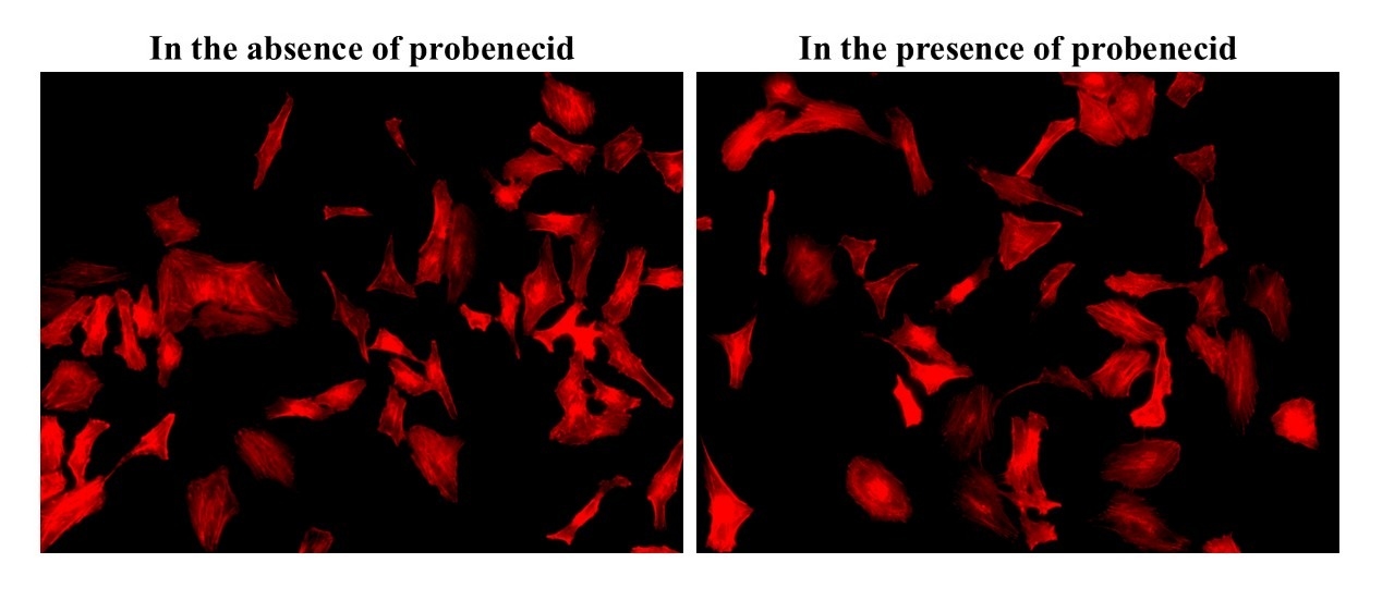
Effect of probenecid (efflux pump inhibitor) on live HeLa cells stained with Actin Lightning™ LC Red (Catalog Number 22705). Live HeLa cells were stained with Actin Lightning™ LC Red in the presence and absence of probenecid. Actin Lightning™ LC Red does not require probenecid and was fully retained in the absence of probenecid.
Actin Lightning™ LC Red also has a significant advantage in that it is a fixable probe. Live cells stained with this label can subsequently be permeabilized and fixed without loss of signal intensity or specificity. This enables compatibility with probes or stains that can only be used on fixed cells, broadening the ability to perform high-plex spatial biology analysis.
Fig. 4
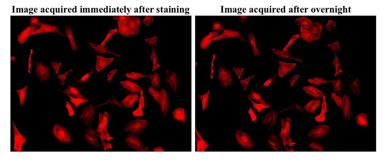
Fixability test for live HeLa cells stained with Actin Lightning™ LC Red (Catalog Number 22705). Live HeLa cells were stained with Actin Lightning™ LC Red followed by fixation using 4% paraformaldehyde. The image shown on left was acquired immediately after staining using Cy5 filter. The image on the right was acquired after overnight following fixation. Actin Lightning™ LC Red does not lose any fluorescence after fixation.
For fixed cell staining of F-actin, phalloidin conjugates have remained a widely used solution. AAT Bioquest improves upon this product class with the release of the Actin Lightning™ iFluor® product line, combining the well-established superior brightness of its iFluor® dye series with a novel, high-affinity, long-lasting F-actin-binding phalloidin. Actin Lightning™ iFluor® Phalloidin Conjugates offer intensely bright fluorescence that is highly specific to F-actin, with a long-lasting signal that can be retained for around a month with minimal loss. These phalloidin conjugates are available in green (FITC), orange (TRITC) and far-red (Cy5) colors, making them compatible with standard fluorescence microscopes. The incorporation of iFluor® dyes also unlocks the possibility of 40+ different color conjugates, enabling high-plex spatial biology applications, such as the study of cell morphology, cytoskeletal organization, and F-actin distribution.
- Highly specific: novel phalloidin results in high-affinity staining with low background
- Powered by iFluor® dyes: conjugates are bright and photostable, with the possibility of 40+ colors
- Multiple colors available: works with standard fluorescence microscopes using FITC/TRITC/Cy5 filters
- Long-lasting signal: can image a month after staining with minimal loss of signal or specificity
Leveraging both a novel, high-affinity phalloidin and superior iFluor® dye performance, Actin Lightning™ iFluor® Phalloidin Conjugates stain F-actin in fixed cells with much higher fluorescence intensity compared to other available labels. In a comparison with another green channel phalloidin conjugate, shown in the figure below, Actin Lightning™ iFluor® 488 Phalloidin Conjugate demonstrated a significantly brighter fluorescence signal intensity under identical experimental conditions, enabling far improved visualization of actin filament structure.
Fig. 5
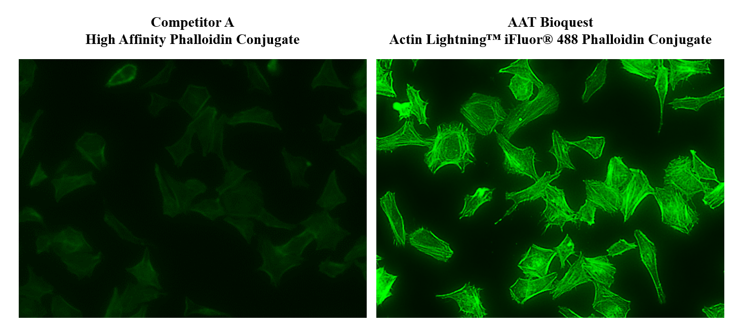
Comparison of F-actin staining in HeLa cells using a commercially available phalloidin conjugate (left) and AAT Bioquest Actin Lightning™ iFluor® 488 Phalloidin Conjugate (right). Images were acquired under identical staining and imaging conditions. Actin Lightning™ iFluor® 488 Phalloidin Conjugate (Catalog Number 22710) produced brighter F-actin staining with higher signal intensity and improved visualization of actin filament structures.
The Actin Lightning™ iFluor® series is supported by AAT Bioquest's high-performance iFluor® dyes, meaning that these conjugates can be available in over 40 different colors. They also benefit from iFluor® dyes' superior photostability and reduced pH sensitivity. These properties offer the flexibility for building large spatial panels for high-plex spatial biology applications.
Fig. 6
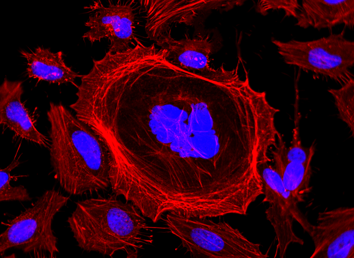
Fluorescence image of HeLa cells stained with Actin Lightning™ iFluor® 570 Phalloidin Conjugate and Nuclear Blue™ DCS1. Cells were fixed with 4% formaldehyde, permeabilized with 0.2% Triton™ X-100, then stained with Actin Lightning™ iFluor® 570 Phalloidin Conjugate (Catalog Number 22716) for F-actin detection and counterstained with Nuclear Blue™ DCS1 (Catalog Number 17548). Images were acquired using Cy3 and DAPI filter sets and overlaid to show F-actin in red and nuclei in blue.
Actin Lightning™ iFluor® Phalloidin Conjugates demonstrate excellent long-term signal stability, with staining detectable even after 30 days, as seen in the figure below. This affords researchers the flexibility for delayed imaging, repeat analysis, high-throughput workflows, and long-term sample review.
Fig. 7
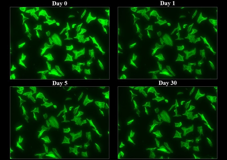
Fluorescence signal stability of Actin Lightning™ iFluor® 488 Phalloidin (Catalog Number 22710, Green) over 30 days. Fixed and permeabilized cells were stained with Actin Lightning™ iFluor® 488 Phalloidin Conjugate and imaged at 0 hour, 1 day, 5 days, and 30 days after staining using a FITC filter set. F-actin staining remained detectable over the 30-day period, supporting long-term signal retention under appropriate conditions.
This document (01.0357.260707r1) was last updated on Tue Jul 14 2026. All trademarks and registered trademarks mentioned herein are the property of their respective owners.
Actin Lightning™ Stains