logo
AAT Bioquest

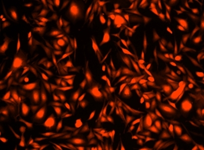
Cell Explorer™ Fixable Live Cell Tracking Kit *Red Fluorescence*

Related catalogs
Our Cell Explorer™ fluorescence imaging kits are a set of tools for labeling cells for fluorescence microscopic investigations of cellular functions. The effective labeling of cells provides a powerful method for studying cellular events in a spatial and temporal context. This particular kit is designed to uniformly label live cells in red fluorescence for the studies that require the fluorescent tag molecules retained inside cells for relatively longer time. The cells can be fixed to retain the imaging pattern. The kit uses a weakly fluorescent dye that carries a cell-retaining moiety. The dye becomes strongly fluorescent upon entering into live cells, and trapped inside live cells to give a stable fluorescence signal for relatively long time. The dye is a hydrophobic compound that easily permeates intact live cells. The labeling process is robust, requiring minimal hands-on time. It can be readily adapted for a wide variety of fluorescence platforms such as microplate assays, immunocytochemistry and flow cytometry. It is useful for a variety of studies, including cell adhesion, chemotaxis, multidrug resistance, cell viability, apoptosis and cytotoxicity. The kit provides all the essential components with an optimized cell-labeling protocol.
Image of HeLa cells stained with Cell Explorer™ Live Cell Tracking Kit in a Costar black wall/clear bottom 96-well plate. Cells were stained with Track It™ Red (Cat#22625) and images were aquired using fluorescence microscope with Texas Red filter set.
Ordering information
Price
Unit size
Catalog Number22625
Quantity
Add to cart
Additional ordering information
Telephone1-800-990-8053
Fax1-800-609-2943
Emailsales@aatbio.com
InternationalSee distributors
Bulk requestInquire
Custom sizeInquire
ShippingStandard overnight for United States, inquire for international
Request quotation
Storage, safety and handling
H-phraseH303, H313, H333
Hazard symbolXN
Intended useResearch Use Only (RUO)
R-phraseR20, R21, R22
UNSPSC12352200
Platform

Flow cytometer

Excitation488 nm laser
Emission610, 20 nm filter
Instrument specification(s)PE-Texas Red channel

Fluorescence microscope

Excitation570 nm
Emission600 nm
Recommended plateBlack wall, clear bottom
Instrument specification(s)Texas Red filter
Components
Example protocol

AT A GLANCE

Protocol summary

  1. Prepare samples
  2. Add 100 µL/well of 1X Track It™ Red working solution
  3. Stain cells at 37°C for 15 minutes to 1 hour
  4. Wash cells
  5. Examine the specimen under microscope with Texas Red filter sets

Important notes
Thaw all the components to room temperature. Centrifuge component A briefly before opening.

PREPARATION OF STOCK SOLUTION

Unless otherwise noted, all unused stock solutions should be divided into single-use aliquots and stored at -20 °C after preparation. Avoid repeated freeze-thaw cycles.

1. Track It™ Red DMSO stock solution (500X):
Add 50 µL DMSO (Component C) into the vial of Component A. Note: The unused portion of the Track It™ Red stock solution should be aliquoted as a single used vials stored at -20 oC. Avoid light and repeated freeze/thaw cycles.

PREPARATION OF WORKING SOLUTION

Dilute 500X Track It™ Red DMSO stock solution into Assay Buffer (Component B). For example, to get a 1X final concentration of Track It™ Red working solution for one 96-well microplate, dilute 20 µL of the Track It™ Red DMSO stock solution into 10 mL of Assay Buffer (Component B). Note: The final concentration of the Track It™ Red working solution should be empirically determined for different cell types and/or experimental conditions. It is recommended to test at the concentrations that are at least over a ten fold range. Note: The working solution should be prepared and used promptly. Keep from light.

For guidelines on cell sample preparation, please visit
https://www.aatbio.com/resources/guides/cell-sample-preparation.html

SAMPLE EXPERIMENTAL PROTOCOL

  1. Remove the cell medium, and add 100 µL/well (for 96 well plate) of 1X Track It™ Red working solution.

  2. Incubate the cells in a 37°C, 5% CO2 incubator for 15 minutes to 1 hour.

  3. Wash cells with Hanks and 20 mM Hepes buffer (HHBS) or an appropriate buffer.

  4. Fill the cell wells with growth medium or fix the cells (optional).

  5. Analyze the cells using a fluorescence microscope with Texas Red filter sets or flow cytometer with 610/20 nm filter (PE-Texas Red channel).
Citations
View all 4 citations: Citation Explorer
The Biological Effects of Interleukin-17A on Adhesion Molecules Expression and Foam Cell Formation in Atherosclerotic Lesions
Authors: Shiotsugu, Shohei and Okinaga, Toshinori and Habu, Manabu and Yoshiga, Daigo and Yoshioka, Izumi and Nishihara, Tatsuji and Ariyoshi, Wataru
Journal: Journal of Interferon & Cytokine Research (2019)
Autophagy proteins are not universally required for phagosome maturation
Authors: Cemma, Marija and Grinstein, Sergio and Brumell, John H
Journal: Autophagy (2016): 1440--1446
Differential detection of tumor cells using a combination of cell rolling, multivalent binding, and multiple antibodies
Authors: Myung, Ja Hye and Gajjar, Khyati A and Chen, Jihua and Molokie, Robert E and Hong, Seungpyo
Journal: Analytical chemistry (2014): 6088--6094
Versatile fabrication of nanoscale sol--gel bioactive glass particles for efficient bone tissue regeneration
Authors: Lei, Bo and Chen, Xiaofeng and Han, Xue and Zhou, Jiaan
Journal: Journal of Materials Chemistry (2012): 16906--16913
References
View all 26 references: Citation Explorer
Requirements, features, and performance of high content screening platforms
Authors: Gough AH, Johnston PA.
Journal: Methods Mol Biol (2007): 41
A pharmaceutical company user's perspective on the potential of high content screening in drug discovery
Authors: Hoffman AF, Garippa RJ.
Journal: Methods Mol Biol (2007): 19
Optimizing the integration of immunoreagents and fluorescent probes for multiplexed high content screening assays
Authors: Giuliano KA., undefined
Journal: Methods Mol Biol (2007): 189
Past, present, and future of high content screening and the field of cellomics
Authors: Taylor DL., undefined
Journal: Methods Mol Biol (2007): 3
G protein-coupled receptor internalization assays in the high-content screening format
Authors: Haasen D, Schnapp A, Valler MJ, Heilker R.
Journal: Methods Enzymol (2006): 121