Products
Services
Resources
Selection Guides
About
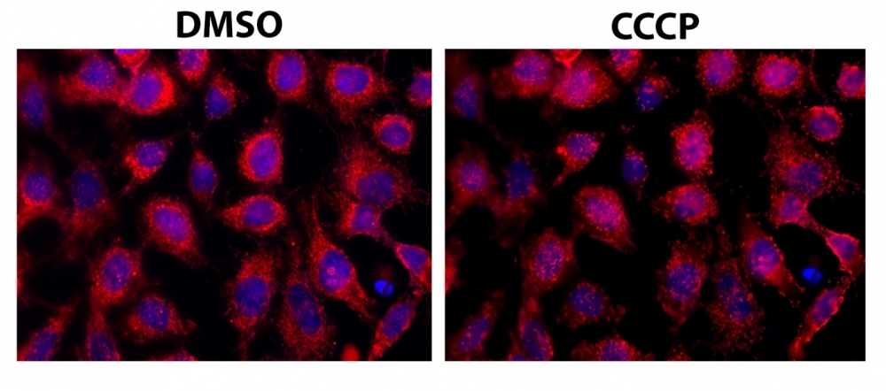
Cell Meter™ Mitochondrial Autophagy Imaging Kit
Red Fluorescence
Mitochondrial autophagy (also called mitophagy) appears to be involved in Alzheimer and Parkinson diseases, mainly induced by the accumulation of depolarized mitochondria. Mitophagy serves as an elimination system that removes dysfunctional mitochondria caused by oxidative stress and DNA damage, causing sequestration into auto phagosome, followed by fusion to lysosome and is degraded. Cell Meter™ Mitochondrial Autophagy Imaging Kit uses Mitophagy Red™ as the mitophagy probe, which enables the very rapid and uniform staining of mitochondria across a wide variety of mammalian cell types and translocate to lysosome upon induction of mitophagy. The Cell Meter™ Mitochondrial Autophagy Imaging Kit provides an excellent tool to be used as an indicator of mitophagy in suspended or attached live cells. The staining pattern of Mitophagy Red™in live cells is stable enough that it provides enough time for studying most live cell dynamic. The assay conditions are compatible with cell culture medium. The excitation/emission of the Mitophagy Red™ probe fits well the widely available Cy3/TRITC filter set and can easily be combined with GFP expressed cell lines if desired.
| Catalog | Size | Price | Quantity |
|---|---|---|---|
| 22998 | 100 Tests | Price |
Usage and storage
| Intended use | Research Use Only (RUO) |
Instrument settings
| Fluorescence microscope | |
| Excitation | Cy3/TRITC filter set |
| Emission | Cy3/TRITC filter set |
| Recommended plate | Black wall/clear bottom |
| Instrument specification(s) | Cy3/TRITC filter set |
Contact us
| Telephone | |
| Fax | |
| sales@aatbio.com | |
| International | See distributors |
| Bulk request | Inquire |
| Custom size | Inquire |
| Technical Support | Contact us |
| Request quotation | Request |
| Purchase order | Send to sales@aatbio.com |
| Shipping | Standard overnight for United States, inquire for international |
Page updated on July 25, 2026