Products
Services
Resources
Selection Guides
About
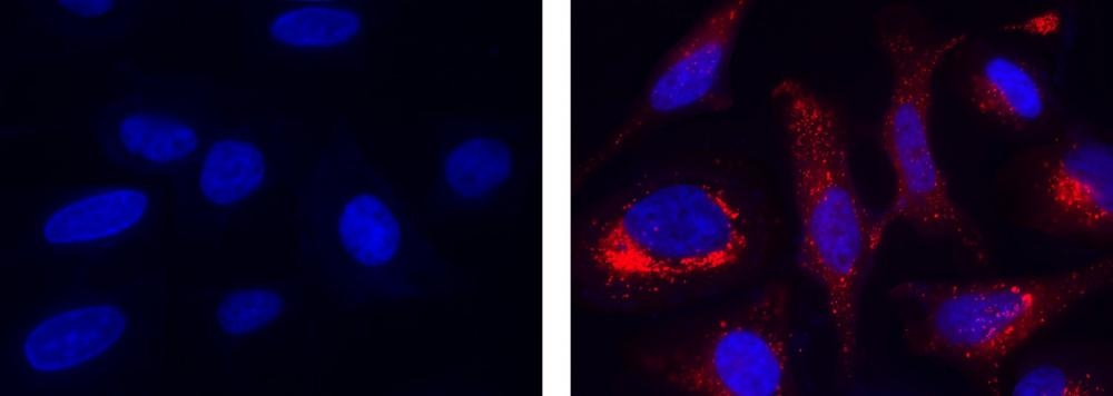
Cell Meter™ Mitochondrial Hydroxyl Radical Detection Kit
Red Fluorescence
The detection of intracellular hydroxyl radical is of central importance to understanding proper cellular redox regulation and the impact of its dysregulation on various pathologies. The hydroxyl radical ('OH) is one of the reactive oxygen species (ROS) highly reactive with other molecules to achieve stability. In general, hydroxyl radical is considered to be a harmful by-product of oxidative metabolism, which can cause molecular damage in living system. It shows an average lifetime of 10-9 nano seconds and can react with nearly every biomolecule such as nuclear DNA, mitochondrial DNA, proteins and membrane lipids. AAT Bioquest's Cell Meter™ Mitochondrial Hydroxyl Radical Detection Kit is optimized for detecting hydroxyl radical in mitochondria. MitoROS™ OH580 is live-cell permeant probe and can rapidly and selectively target hydroxyl radical in live cells. It generates red fluorescence when it reacts with 'OH, and can be easily read. Cell Meter™ Mitochondrial Hydroxyl Radical Detection Kit provides a sensitive fluorimetric probe to detect OH' in live cells with one hour incubation. This kit can be used for fluorescence microplate readers and fluorescence microscopy applications.
| Catalog | Size | Price | Quantity |
|---|---|---|---|
| 16055 | 200 Tests | Price |

Spectral properties
| Excitation (nm) | 576 |
| Emission (nm) | 598 |
Storage, safety and handling
| H-phrase | H303, H313, H333 |
| Hazard symbol | XN |
| Intended use | Research Use Only (RUO) |
| R-phrase | R20, R21, R22 |
| UNSPSC | 12352200 |
Instrument settings
| Fluorescence microscope | |
| Excitation | Cy3/TRITC filter set |
| Emission | Cy3/TRITC filter set |
| Recommended plate | Black wall/clear bottom |
| Fluorescence microplate reader | |
| Excitation | 540 nm |
| Emission | 590 nm |
| Cutoff | 570 nm |
| Recommended plate | Black wall/clear bottom |
| Instrument specification(s) | Bottom read mode |
Contact us
| Telephone | |
| Fax | |
| sales@aatbio.com | |
| International | See distributors |
| Bulk request | Inquire |
| Custom size | Inquire |
| Technical Support | Contact us |
| Request quotation | Request |
| Purchase order | Send to sales@aatbio.com |
| Shipping | Standard overnight for United States, inquire for international |
Page updated on June 27, 2026