Cell Meter™ NIR Mitochondrion Membrane Potential Assay Kit *Optimized for Microplate Reader*
Ordering information
Price | |
Catalog Number | |
Unit Size | |
Quantity |
Additional ordering information
Telephone | 1-800-990-8053 |
Fax | 1-800-609-2943 |
sales@aatbio.com | |
International | See distributors |
Bulk request | Inquire |
Custom size | Inquire |
Shipping | Standard overnight for United States, inquire for international |
Storage, safety and handling
H-phrase | H303, H313, H333 |
Hazard symbol | XN |
Intended use | Research Use Only (RUO) |
R-phrase | R20, R21, R22 |
UNSPSC | 12352200 |
Alternative formats
Cell Meter™ NIR Mitochondrion Membrane Potential Assay Kit *Optimized for Flow Cytometry* |
Related products
Overview | ![]() ![]() |
See also: Mitochondrial Membrane Potential
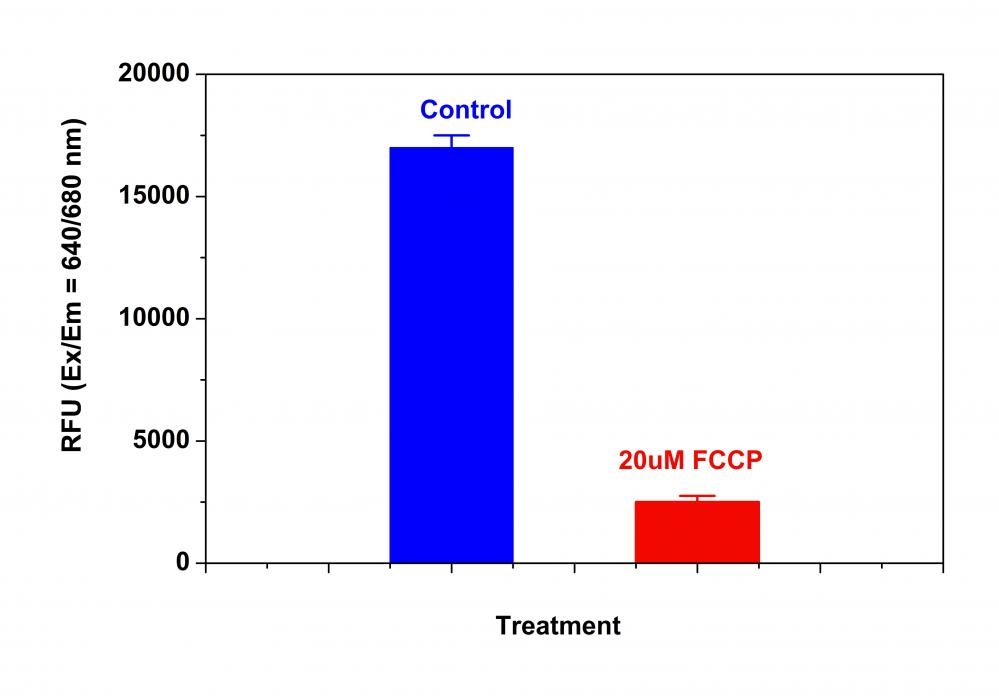
Our Cell Meter™ assay kits are a set of tools for monitoring cell viability. There are a variety of parameters that can be used for monitoring cell viability. This particular kit is designed to monitor cell apoptosis through measuring the loss of the mitochondrial membrane potential. The collapse of mitochondrial membrane potential coincides with the opening of the mitochondrial permeability transition pores, leading to the release of cytochrome C into the cytosol, which in turn triggers other downstream events in the apoptotic cascade. Our Cell Meter™ NIR Mitochondria Membrane Potential Detection Kit provides all the essential components with an optimized assay method for the detection of apoptosis in cells with the loss of mitochondrial membrane potential. This fluorometric assay is based on the detection of the mitochondrial membrane potential in cells by our proprietary cationic MitoLite NIR™ dye. In normal cells, MitoLite NIR™ accumulates primarily in mitochondria, however, in apoptotic cells, MitoLite NIR™ staining intensity decreases. Cells stained with MitoLite NIR™ can be monitored fluorimetrically at 660-680 nm with excitation of 620-640 nm. The kit can be used for screening of apoptosis activators and inhibitors. The assay can be performed in a convenient 96-well and 384-well fluorescence microtiter-plate format.
Platform
Fluorescence microplate reader
Excitation | 640 nm |
Emission | 680 nm |
Cutoff | 665 nm |
Recommended plate | Black wall/clear bottom |
Instrument specification(s) | Bottom read mode |
Components
Example protocol
AT A GLANCE
Protocol summary
- Prepare cells
- Add test compounds
- Add MitoLite™ NIR working solution (100 µL/well/ 96-well plate or 25 µL/well/384-well plate)
- Incubate at 37°C, 5% CO2 incubator for 30 - 60 minutes
- Add Assay Buffer B (50 µL/well/96-well plate or 12.5 µL/well/384-well plate)
- Monitor fluorescence intensity (Bottom read mode) at Ex/Em = 640/680 nm (Cutoff = 665 nm)
Important notes
Thaw all the kit components at room temperature before starting the experiment.
PREPARATION OF WORKING SOLUTION
Add 50 µL of 200X MitoLite™ NIR (Component A) into 10 mL of Assay Buffer A (Component B) and mix well to make MitoLite™ NIR working solution. Protect from light.
For guidelines on cell sample preparation, please visit
https://www.aatbio.com/resources/guides/cell-sample-preparation.html
SAMPLE EXPERIMENTAL PROTOCOL
- Treat cells with test compounds for a desired period of time to induce apoptosis and set up parallel control experiments.
For Negative Control: Treat cells with vehicle only.
For Positive Control: Treat cells with FCCP or CCCP at 5 - 50 µM in a 37°C, 5% CO2 incubator for 15 to 30 minutes. Note: CCCP or FCCP can be added simultaneously with MitoLite™ NIR. To get the best result, titration of the CCCP or FCCP may be required for each individual cell line. - Remove the cell medium before adding MitoLite™ NIR working solution. Note: It is important to remove the cell medium before adding MitoLite™ NIR working solution.
- Add 100 µL/well/96-well plate or 25 µL/well/384-well plate of MitoLite™ NIR working solution into the cell plate.
- Incubate the plate in a 37°C, 5% CO2 incubator for 30 - 60 minutes, protected from light. Note: The appropriate incubation time depends on the individual cell type and cell concentration used. Optimize the incubation time for each experiment.
- Add 50 µL/well/96-well plate or 12.5 µL/well/384-well plate of Assay Buffer B (Component C) into the cell plate before monitoring the fluorescence signal. Note: DO NOT wash the cells after loading. For non-adherent cells, it is recommended to centrifuge cell plates at 800 rpm for 2 minutes with brake off after adding Assay Buffer B (Component C).
- Monitor the fluorescence intensity with a fluorescence microplate reader (bottom read mode) at Ex/Em = 640/680 nm (Cutoff = 665 nm) either using the endpoint mode or using the kinetic mode 10 to 30 minutes after adding Assay Buffer B (Component C).
Images

Figure 1. The decrease in MitoLite™ NIR fluorescence with the addition of FCCP in HeLa cells. HeLa cells were loaded with MitoLite™ NIR alone or in the presence of 20 µM FCCP for 15 minutes. The fluorescence intensity of MitoLite™ NIR was measured 30 minutes after adding assay buffer with a FlexStation™ microplate reader (Molecular Devices) at Ex/Em = 640/680 nm (Cutoff = 665 nm, bottom read mode).
References
View all 91 references: Citation Explorer
Safranine O as a fluorescent probe for mitochondrial membrane potential studied on the single particle level and in suspension
Authors: Perevoshchikova IV, Sorochkina AI, Zorov DB, Antonenko YN.
Journal: Biochemistry (Mosc) (2009): 663
Authors: Perevoshchikova IV, Sorochkina AI, Zorov DB, Antonenko YN.
Journal: Biochemistry (Mosc) (2009): 663
Determination of high mitochondrial membrane potential in spermatozoa loaded with the mitochondrial probe 5,5',6,6'-tetrachloro-1,1',3,3'-tetraethylbenzimidazolyl-carbocyanine iodide (JC-1) by using fluorescence-activated flow cytometry
Authors: Guthrie HD, Welch GR.
Journal: Methods Mol Biol (2008): 89
Authors: Guthrie HD, Welch GR.
Journal: Methods Mol Biol (2008): 89
The mitochondrial membrane potential and Ca2+ oscillations in smooth muscle
Authors: Chalmers S, McCarron JG.
Journal: J Cell Sci (2008): 75
Authors: Chalmers S, McCarron JG.
Journal: J Cell Sci (2008): 75
Computer-assisted live cell analysis of mitochondrial membrane potential, morphology and calcium handling
Authors: Koopman WJ, Distelmaier F, Esseling JJ, Smeitink JA, Willems PH.
Journal: Methods (2008): 304
Authors: Koopman WJ, Distelmaier F, Esseling JJ, Smeitink JA, Willems PH.
Journal: Methods (2008): 304
How DASPMI reveals mitochondrial membrane potential: fluorescence decay kinetics and steady-state anisotropy in living cells
Authors: Ramadass R, Bereiter-Hahn J.
Journal: Biophys J (2008): 4068
Authors: Ramadass R, Bereiter-Hahn J.
Journal: Biophys J (2008): 4068
Life cell quantification of mitochondrial membrane potential at the single organelle level
Authors: Distelmaier F, Koopman WJ, Testa ER, de Jong AS, Swarts HG, Mayatepek E, Smeitink JA, Willems PH.
Journal: Cytometry A (2008): 129
Authors: Distelmaier F, Koopman WJ, Testa ER, de Jong AS, Swarts HG, Mayatepek E, Smeitink JA, Willems PH.
Journal: Cytometry A (2008): 129
Effects of eprosartan on mitochondrial membrane potential and H2O2 levels in leucocytes in hypertension
Authors: Labios M, Martinez M, Gabriel F, Guiral V, Ruiz-Aja S, Beltran B, Munoz A.
Journal: J Hum Hypertens (2008): 493
Authors: Labios M, Martinez M, Gabriel F, Guiral V, Ruiz-Aja S, Beltran B, Munoz A.
Journal: J Hum Hypertens (2008): 493
Mitochondrial membrane potential in axons increases with local nerve growth factor or semaphorin signaling
Authors: Verburg J, Hollenbeck PJ.
Journal: J Neurosci (2008): 8306
Authors: Verburg J, Hollenbeck PJ.
Journal: J Neurosci (2008): 8306
Evaluation of sperm mitochondrial membrane potential by JC-1 fluorescent staining and flow cytometry
Authors: Xia XY, Wu YM, Hou BS, Yang B, Pan LJ, Shi YC, Jin BF, Shao Y, Cui YX, Huang YF.
Journal: Zhonghua Nan Ke Xue (2008): 135
Authors: Xia XY, Wu YM, Hou BS, Yang B, Pan LJ, Shi YC, Jin BF, Shao Y, Cui YX, Huang YF.
Journal: Zhonghua Nan Ke Xue (2008): 135
Cyclosporin A-induced oxidative stress is not the consequence of an increase in mitochondrial membrane potential
Authors: van der Toorn M, Kauffman HF, van der Deen M, Slebos DJ, Koeter GH, Gans RO, Bakker SJ.
Journal: Febs J (2007): 3003
Authors: van der Toorn M, Kauffman HF, van der Deen M, Slebos DJ, Koeter GH, Gans RO, Bakker SJ.
Journal: Febs J (2007): 3003
Application notes
A Library of Well-Defined and Water-Soluble Poly(alkyl phosphonate)s with Adjustable Hydrolysis
A New Red Fluorescent & Robust Screen Quest™ Rhod-4™ Ca2+Indicator for Screening GPCR & Ca2+ Channel Targets
A New Robust No-Wash FLIPR Calcium Assay Kit for Screening GPCR and Calcium Channel Targets
A Non-Radioactive Photometric Assay for Glucose Uptake in Insulin-Responsive 3T3-L1 Adipocytes
A Novel Fluorescent Probe for Imaging and Detecting Hydroxyl Radical in Living Cells