Cell Meter™ JC-10 Mitochondrion Membrane Potential Assay Kit *Optimized for Microplate Assays*
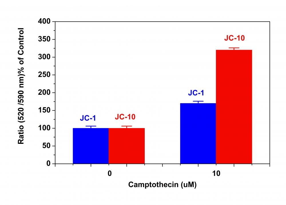
Although JC-1 is widely used in many labs, its poor water solubility makes it hard to use for some applications. Even at 1 µM concentration, JC-1 tends to precipitate in aqueous buffer. JC-10 has been developed to be a superior alternative to JC-1 where high dye concentration is desired. Compared to JC-1, our JC-10 has much better water solubility. JC-10 is capable of entering selectively into mitochondria, and changes reversibly its color from green to orange as membrane potentials increase. This property is due to the reversible formation of JC-10 aggregates upon membrane polarization that causes shifts in emitted light from 520 nm (i.e., emission of JC-10 monomeric form) to 570 nm (i.e., emission of J-aggregate). When excited at 490 nm, the color of JC-10 changes reversibly from green to greenish orange as the mitochondrial membrane becomes more polarized. This Cell Meter™ JC-10 Mitochondrial Membrane Potential Assay Kit enable you to monitor mitochondrial membrane potential changes using a simple microplate reader while all the other commercial JC-1 assay kits require the use of a flow cytometer. Our kit provides the most robust method to monitor mitochondrial membrane potential changes, and can be readily used for screening a large compound library.
![Campotothecin-induced mitochondria membrane potential changes were measured with JC-10™ and JC-1 in Jurkat cells. After Jurkat cells were treated with camptothecin (10 µM) for 4 hours, JC-1 and JC-10™ dye working solutions were added to the wells and incubated for 30 minutes. The fluorescence intensities for both J-aggregates (highlighted in blue) and monomeric forms (highlighted in red) of JC-1 and JC-10™ were measured at Ex/Em = 490/525 nm (Cutoff = 515 nm) and 490/590 nm (Cutoff = 570 nm) with NOVOstar microplate reader (BMG Labtech).](/_next/image?url=https%3A%2F%2Fimages.aatbio.com%2Fproducts%2Ffigures-and-data%2Fcell-meter-jc-10-mitochondrion-membrane-potential-assay-kit-optimized-for-microplate-assays%2Ffigure-for-cell-meter-jc-10-mitochondrion-membrane-potential-assay-kit-optimized-for-microplate-assays_2YJlW.jpg&w=640&q=75)
![Campotothecin-induced mitochondria membrane potential changes were measured with JC-10™ and JC-1 in Jurkat cells. After Jurkat cells were treated with camptothecin (10 µM) for 4 hours, JC-1 and JC-10™ dye working solutions were added to the wells and incubated for 30 minutes. The fluorescence intensities for both J-aggregates (highlighted in blue) and monomeric forms (highlighted in red) of JC-1 and JC-10™ were measured at Ex/Em = 490/525 nm (Cutoff = 515 nm) and 490/590 nm (Cutoff = 570 nm) with NOVOstar microplate reader (BMG Labtech).](/_next/image?url=https%3A%2F%2Fimages.aatbio.com%2Fproducts%2Ffigures-and-data%2Fcell-meter-jc-10-mitochondrion-membrane-potential-assay-kit-optimized-for-microplate-assays%2Ffigure-for-cell-meter-jc-10-mitochondrion-membrane-potential-assay-kit-optimized-for-microplate-assays_2YJlW.jpg&w=128&q=25)
Example protocol
AT A GLANCE
Protocol summary
- Prepare cells
- Add test compounds
- Add JC-10 dye-working solution (50 µL/well/96-well plate or 12.5 µL/well/384-well plate)
- Incubate at 37°C, 5% CO2 incubator for 30 to 60 minutes
- Add Assay Buffer B (50 µL/well/96-well plate or 12.5 µL/well/384-well plate)
- Monitor fluorescence intensities (bottom read mode) at Ex/Em = 490/525 nm (Cutoff = 515 nm) and 540/590 nm (Cutoff = 570 nm)
Important notes
Thaw all the kit components at room temperature before starting the experiment.
PREPARATION OF WORKING SOLUTION
Add 50 µL of 100X JC-10 (Component A) into 5 mL of Assay Buffer A (Component B) and mix well to make JC-10 dye-working solution. Protect from light.
For guidelines on cell sample preparation, please visit
https://www.aatbio.com/resources/guides/cell-sample-preparation.html
SAMPLE EXPERIMENTAL PROTOCOL
- Treat cells by adding 10 µL of 10X test compounds (96-well plate) or 5 µL of 5X test compounds (384-plate) into the desired buffer (such as PBS or HHBS). Note: It is not necessary to wash cells before adding compound. However, if tested compounds are serum sensitive, growth medium and serum factors can be aspirated away before adding compounds. Add the same volume of HHBS into the wells (such as 90 µL for a 96-well plate or 20 µL for a 384-well plate) after aspiration. Alternatively, cells can be grown in serum-free media.
- Incubate the cell plate at room temperature or in a 37°C, 5% CO2 incubator for at least 15 minutes or a desired period of time (for Jurkat cells, 4 - 6 hours with camptothecin or 3 - 5 hours with staurosporine treatment) to induce apoptosis.
- Add 50 µL/well (96-well plate) or 12.5 µL/well (384-well plate) of JC-10 dye-working solution into the cell plate.
- Incubate the plate in a 37°C, 5% CO2 incubator for 30 - 60 minutes, protected from light. Note: The appropriate incubation time depends on the individual cell type and cell concentration used. Optimize the incubation time for each experiment.
- Add 50 µL/well (96-well plate) or 12.5 µL/well (384-well plate) of Assay Buffer B (Component C) into JC-10 dye-working solution plate before reading the fluorescence intensity. Note: DO NOT wash the cells after loading. For non-adherent cells, it is recommended to centrifuge cell plates at 800 rpm for 2 minutes with brake off after adding Assay Buffer B (Component C).
- Monitor the fluorescence intensities with a fluorescence microplate reader (bottom read mode) at Ex/Em = 490/525 nm (Cutoff = 515 nm) and 540/590 nm (Cutoff = 570 nm) for ratio analysis.
Spectrum
Citations
View all 92 citations: Citation Explorer
Mitochondria of Porcine Oocytes Synthesize Melatonin, Which Improves Their In Vitro Maturation and Embryonic Development
Authors: Zhu, Tianqi and Yan, Laiqing and Deng, Shoulong and Ma, Wenkui and Xia, Fan and Wang, Likai and Ma, Xiao and Li, Guangdong and Shen, Zixia and Wang, Yiwei and others,
Journal: Antioxidants (2024): 814
Authors: Zhu, Tianqi and Yan, Laiqing and Deng, Shoulong and Ma, Wenkui and Xia, Fan and Wang, Likai and Ma, Xiao and Li, Guangdong and Shen, Zixia and Wang, Yiwei and others,
Journal: Antioxidants (2024): 814
Astrocyte Mitochondria Are a Sensitive Target of PCB52 and its Human-Relevant Metabolites
Authors: Paranjape, Neha and Strack, Stefan and Lehmler, Hans-Joachim and Doorn, Jonathan A
Journal: ACS Chemical Neuroscience (2024)
Authors: Paranjape, Neha and Strack, Stefan and Lehmler, Hans-Joachim and Doorn, Jonathan A
Journal: ACS Chemical Neuroscience (2024)
Green synthesis and characterization of AgNPs, liposomal loaded AgNPs and ZnPcS4 photosensitizer for enhanced photodynamic therapy effects in MCF-7 breast cancer cells
Authors: Chota, Alexander and Abrahamse, Heidi and George, Blassan P
Journal: Photodiagnosis and Photodynamic Therapy (2024): 104252
Authors: Chota, Alexander and Abrahamse, Heidi and George, Blassan P
Journal: Photodiagnosis and Photodynamic Therapy (2024): 104252
Chlorogenic Acid as a Potential Therapeutic Agent for Cholangiocarcinoma
Authors: Liang, Jiabao and Wen, Tong and Zhang, Xiaojian and Luo, Xiaoling
Journal: Pharmaceuticals (2024): 794
Authors: Liang, Jiabao and Wen, Tong and Zhang, Xiaojian and Luo, Xiaoling
Journal: Pharmaceuticals (2024): 794
Effect of autologous platelet-rich plasma on the fertility and quality of cryopreserved buffalo bull semen: a comparative study using OptiXcell{\textregistered} and tris egg yolk extenders
Authors: Salama, Maha S and Ashour, Mohey A and Taher, Ehab S and Rashed, Fatema and Ibrahim, Ibrahim M and El-Nablaway, Mohammad and Ibrahim, Ateya Megahed and Mihaela, Ostan and Olga, Rada and Mohammed, Nourelhuda A and others,
Journal: BMC Veterinary Research (2024): 1--10
Authors: Salama, Maha S and Ashour, Mohey A and Taher, Ehab S and Rashed, Fatema and Ibrahim, Ibrahim M and El-Nablaway, Mohammad and Ibrahim, Ateya Megahed and Mihaela, Ostan and Olga, Rada and Mohammed, Nourelhuda A and others,
Journal: BMC Veterinary Research (2024): 1--10