Products
Services
Resources
Selection Guides
About
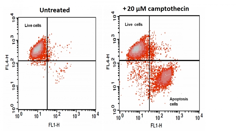
Cell Meter™ Nuclear Apoptosis Assay Kit
Green Fluorescence Optimized for Flow Cytometry
Our Cell Meter™ assay kits are a set of tools for monitoring cell viability. There are a variety of parameters that can be used for monitoring cell viability. This particular kit is designed to monitor cell apoptosis through measuring the apoptotic chromatin condensation. The compacted chromatin of apoptotic cells binds higher amounts of nuclear dye compared to the healthy cells. Our Cell Meter™ Nuclear Apoptosis Assay Kit provides all the essential components with an optimized assay method for the detection of apoptosis in cells with condensed nuclei. This fluorometric assay is based on the detection of the DNA contents in cells using our proprietary non-fluorescent dye that becomes strongly fluorescent upon binding to DNA. In normal cells, Nuclear Green™ is not cell permeable, however, in apoptotic cells, Cells with compromised plasma membranes or with impaired/no cell metabolism are unable to prevent the dye from entering the cell. Once inside the cell, the dyes bind to intracellular DNA producing highly fluorescent complexes which identify the cells as non-viable cells. The staining with Nuclear Green; DCS1 can be measured using a flow cytometer (FL1 channel) or fluorescence microscope (FITC filter set). The kit can be used with our other apoptosis reagents, such as Our Cell Meter™ NIR Mitochondria Membrane Potential Detection Kit (Cat# 22802), for multi-parametric study of cell viability and apoptosis. The kit is optimized for screening of apoptosis activators and inhibitors.
| Catalog | Size | Price | Quantity |
|---|---|---|---|
| 22811 | 100 Tests | Price |

Spectral properties
| Excitation (nm) | 503 |
| Emission (nm) | 527 |
Usage and storage
| Intended use | Research Use Only (RUO) |
Instrument settings
| Flow cytometer | |
| Excitation | 488 nm laser |
| Emission | 530/30 nm filter |
| Instrument specification(s) | FITC channel |
Contact us
| Telephone | |
| Fax | |
| sales@aatbio.com | |
| International | See distributors |
| Bulk request | Inquire |
| Custom size | Inquire |
| Technical Support | Contact us |
| Request quotation | Request |
| Purchase order | Send to sales@aatbio.com |
| Shipping | Standard overnight for United States, inquire for international |
Page updated on July 16, 2026