Products
Services
Resources
Selection Guides
About
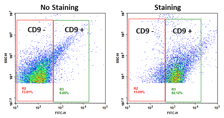
Cell Navigator® Flow Cytometric Exosome Staining Kit
Exosomes, nanoscale (30-150 nm) membrane-bound vesicles secreted by cells, are central to intercellular communication. They transfer proteins, lipids, and nucleic acids, including RNA species, between cells, playing a pivotal role in physiological and pathological processes such as immune modulation, disease progression, and genetic information exchange. This makes exosomes key targets in therapeutic and diagnostic research. However, challenges such as dye aggregation and the absence of bright, specific probes have hindered the reliable detection and analysis of exosomes. The Cell Navigator® Flow Cytometric Exosome Staining Kit addresses these challenges by enabling the detection of CD9-positive exosomes in purified or bead-bound samples. It utilizes a fluorescent dye-conjugated CD9 marker for targeted visualization, providing a straightforward staining protocol for rapid and accurate exosome detection in diverse sample matrices, including cell culture supernatants, biological fluids, and isolated exosome preparations. To ensure effective detection and analysis, it is essential to enrich the cell culture media for exosomes before staining. This can be achieved through efficient methods such as the ReadiPrep™ Exosome Isolation Kit (Cat No. 60204) or traditional ultracentrifugation.
| Catalog | Size | Price | Quantity |
|---|---|---|---|
| 22426 | 100 Tests | Price |

Spectral properties
| Absorbance (nm) | 488 |
| Correction factor (280 nm) | 0.35 |
| Extinction coefficient (cm -1 M -1) | 73000 |
| Excitation (nm) | 491 |
| Emission (nm) | 516 |
| Quantum yield | 0.92 |
Usage and storage
| Intended use | Research Use Only (RUO) |
| Storage | Refrigerated (2-8 °C); Minimize light exposure |
Instrument settings
| Flow cytometer | |
| Excitation | 488 nm Laser |
| Emission | 530/30 nm Filter |
| Instrument specification(s) | FITC Channel |
Contact us
| Telephone | |
| Fax | |
| sales@aatbio.com | |
| International | See distributors |
| Bulk request | Inquire |
| Custom size | Inquire |
| Technical Support | Contact us |
| Request quotation | Request |
| Purchase order | Send to sales@aatbio.com |
| Shipping | Standard overnight for United States, inquire for international |
Page updated on July 14, 2026