logo
AAT Bioquest

Cy7® goat anti-rabbit IgG (H+L) *Cross Adsorbed*

AAT Bioquest's anti-rabbit secondary antibodies are affinity-purified antibodies with well-characterized specificity for rabbit immunoglobulins and are useful in the detection, sorting or purification of its specified target. This Cy7-labeled secondary antibody was prepared using AAT Bioquest's proprietary labeling technology. It demonstrated much brighter signal compared to the similar Cy7 goat anti-rabbit IgG antibodies from other commercial sources, thus can significantly increase assay sensitivities. Secondary antibodies offer increased versatility enabling users to use many detection systems (e.g. HRP, AP, fluorescence). They can also provide greater sensitivity through signal amplification as multiple secondary antibodies can bind to a single primary antibody.
Spectrum

Product family

NameExcitation (nm)Emission (nm)Extinction coefficient (cm -1 M -1)Quantum yieldCorrection Factor (260 nm)Correction Factor (280 nm)
Cy7® goat anti-mouse IgG (H+L)7567792500000.30.050.036
Cy7® goat anti-mouse IgG (H+L) *Cross Adsorbed*7567792500000.30.050.036
FITC goat anti-rabbit IgG (H+L)491516730000.92-0.35
Cy3® goat anti-rabbit IgG (H+L)55556915000010.1510.070.073
Cy5® goat anti-rabbit IgG (H+L)65167025000010.271, 0.420.020.03
FITC goat anti-rabbit IgG (H+L) *Cross Adsorbed*491516730000.92-0.35
Cy3® goat anti-rabbit IgG (H+L) *Cross Adsorbed*55556915000010.1510.070.073
Cy5® goat anti-rabbit IgG (H+L) *Cross Adsorbed*65167025000010.271, 0.420.020.03
XFD350 goat anti-rabbit IgG (H+L) *Cross Adsorbed, XFD350 Same Structure to Alexa Fluor™ 350*34344119000-0.250.19
XFD488 goat anti-rabbit IgG (H+L) *Cross Adsorbed, XFD488 Same Structure to Alexa Fluor™ 488*499520730000.9210.30.11
XFD594 goat anti-rabbit IgG (H+L) *Cross Adsorbed, XFD594 Same Structure to Alexa Fluor™ 594*590618920000.6610.430.56
Show More (2)

Citations

View all 4 citations: Citation Explorer
Overexpression of CXCR2 predicts poor prognosis in patients with colorectal cancer.
Authors: Zhao, Jingkun and Ou, Baochi and Feng, Hao and Wang, Puxiongzhi and Yin, Shuai and Zhu, Congcong and Wang, Shenjie and Chen, Chun and Zheng, Minhua and Zong, Yaping and others, undefined
Journal: Oncotarget (2017)
Cadherin-12 enhances proliferation in colorectal cancer cells and increases progression by promoting EMT
Authors: Ma, Junjun and Zhao, Jingkun and Lu, Jun and Wang, Puxiongzhi and Feng, Hao and Zong, Yaping and Ou, Baochi and Zheng, Minhua and Lu, Aiguo
Journal: Tumor Biology (2016): 1--12
Transplantation of RADA16-BDNF peptide scaffold with human umbilical cord mesenchymal stem cells forced with CXCR4 and activated astrocytes for repair of traumatic brain injury
Authors: Shi, W and Huang, CJ and Xu, XD and Jin, GH and Huang, RQ and Huang, JF and Chen, YN and Ju, SQ and Wang, Y and Shi, YW and others, undefined
Journal: Acta Biomaterialia (2016): 247--261
The migration and differentiation of hUC-MSCsCXCR4/GFP encapsulated in BDNF/chitosan scaffolds for brain tissue engineering
Authors: Huang, Chuanjun and Zhao, Longxiang and Gu, Jun and Nie, Dekang and Chen, Yinan and Zuo, Hao and Huan, Wei and Shi, Jinlong and Chen, Jian and Shi, Wei
Journal: Biomedical Materials (2016): 035004

References

View all 191 references: Citation Explorer
Antiprothrombin antibodies in a patient with secondary antiphospholipid syndrome and bleeding
Authors: Gonzalez Leon R, Garcia Hern and ez FJ, Castillo Palma MJ, Sanchez Roman J.
Journal: Med Clin (Barc) (2011): 668
Assessment of EGFR/HER2 dimerization by FRET-FLIM utilizing Alexa-conjugated secondary antibodies in relation to targeted therapies in cancers
Authors: Waterhouse BR, Gijsen M, Barber PR, Tullis ID, Vojnovic B, Kong A.
Journal: Oncotarget (2011): 728
Falsely elevated tacrolimus levels caused by immunoassay interference secondary to beta-galactosidase antibodies in an infected liver transplant recipient
Authors: Knorr JP, Grewal KS, Balasubramanian M, Young N, Zaki R, Khanmoradi K, Araya V, Ortiz J.
Journal: Pharmacotherapy (2010): 954
Prevalence of anti-Epstein-Barr virus antibodies in children and adolescents with secondary immunodeficiency
Authors: Buckova A., undefined
Journal: Epidemiol Mikrobiol Imunol (2010): 133
Analysis of the effectiveness of reused primary and secondary antibodies in Western blotting analysis
Authors: Boonrod K, Roth B, Leong Ngar S, Krczal G.
Journal: Anal Biochem (2010): 124
Page updated on December 13, 2024

Ordering information

Price
Unit size
Catalog Number16880
Quantity
Add to cart

Additional ordering information

Telephone1-800-990-8053
Fax1-800-609-2943
Emailsales@aatbio.com
InternationalSee distributors
Bulk requestInquire
Custom sizeInquire
Technical SupportContact us
Purchase orderSend to sales@aatbio.com
ShippingStandard overnight for United States, inquire for international
Request quotation

Physical properties

Molecular weight

~150000

Solvent

Water

Spectral properties

Correction Factor (260 nm)

0.05

Correction Factor (280 nm)

0.036

Correction Factor (482 nm)

0.0005

Correction Factor (565 nm)

0.0193

Correction Factor (650 nm)

0.165

Extinction coefficient (cm -1 M -1)

250000

Excitation (nm)

756

Emission (nm)

779

Quantum yield

0.3

Storage, safety and handling

H-phraseH303, H313, H333
Hazard symbolXN
Intended useResearch Use Only (RUO)
R-phraseR20, R21, R22

Storage

Freeze (< -15 °C); Minimize light exposure
UNSPSC12171501
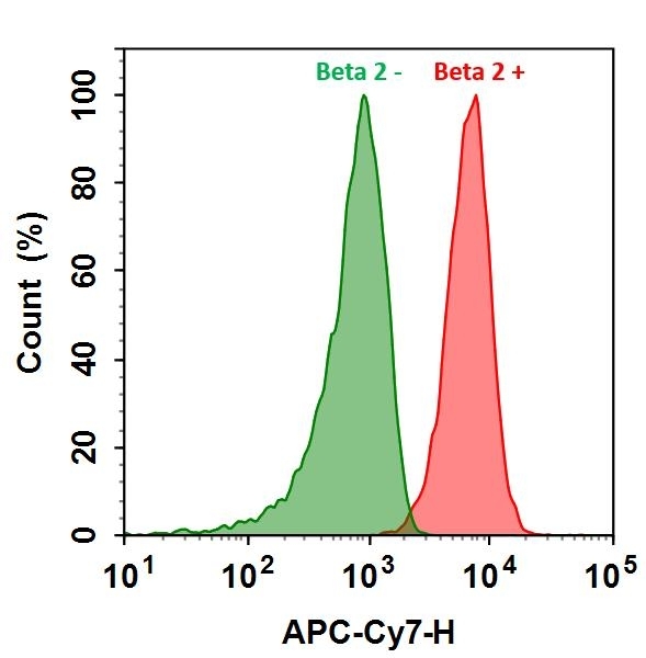
HL-60 cells were incubated with (Red, +) or without (Green, -) Anti-beta 2 rabbit antibody (Beta 2), followed by Cy7&reg; goat anti-rabbit IgG conjugate. The fluorescence signal was monitored using ACEA NovoCyte flow cytometer in APC-Cy7 channel.