Products
Services
Resources
Selection Guides
About
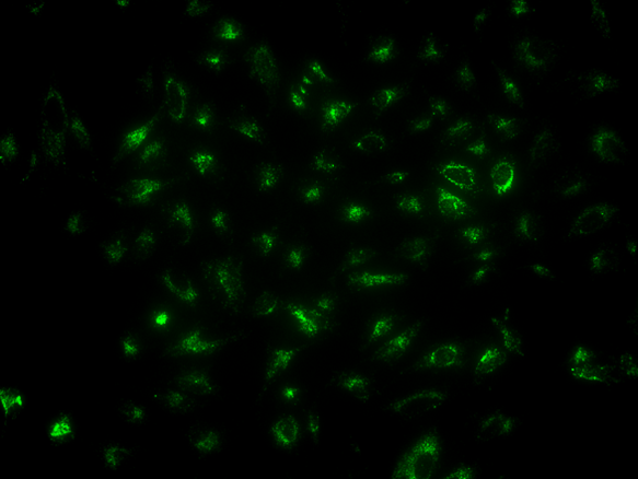
LysoBrite™ Green DND-26
LysoBrite™ Green DND-26 is chemically the same as the LysoTracker® Green DND-26 used for labeling and tracking acidic organelles in live cells (LysoTracker® is the trademark of ThermoFisher). It has good selectivity for acidic organelles. The LysoBrite™ probes consist of a fluorophore linked to a weak base that is only partially protonated at neutral pH, allowing them to freely permeate cell membranes to label live cells.
| Catalog | Size | Price | Quantity |
|---|---|---|---|
| 22648 | 500 Tests | Price |

Physical properties
| Molecular weight | 362.23 |
| Solvent | DMSO |
Spectral properties
| Excitation (nm) | 501 |
| Emission (nm) | 509 |
Storage, safety and handling
| H-phrase | H303, H313, H333 |
| Hazard symbol | XN |
| Intended use | Research Use Only (RUO) |
| R-phrase | R20, R21, R22 |
| Storage | Freeze (< -15 °C); Minimize light exposure |
| UNSPSC | 12171501 |
Instrument settings
| Flow cytometer | |
| Excitation | 488 nm laser |
| Emission | 530/30 nm filter |
| Instrument specification(s) | FITC channel |
| Fluorescence microscope | |
| Excitation | FITC filter set |
| Emission | FITC filter set |
| Recommended plate | Black wall/clear bottom |
Contact us
| Telephone | |
| Fax | |
| sales@aatbio.com | |
| International | See distributors |
| Bulk request | Inquire |
| Custom size | Inquire |
| Technical Support | Contact us |
| Request quotation | Request |
| Purchase order | Send to sales@aatbio.com |
| Shipping | Standard overnight for United States, inquire for international |
Page updated on July 6, 2026