Products
Services
Resources
Selection Guides
About
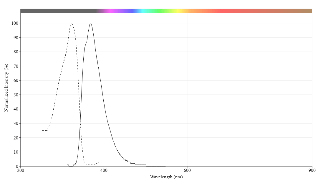
mFluor™ UVB 370 SE
mFluor™ UVB 370 SE is a UV excitable fluorophore (Ex: 320 nm) with unique spectral properties that expands multicolor flow cytometry into the deep UV range for advanced panel design.
- Unique UV Spectral Window: Excitation at ~320 nm and emission at ~370 nm provides a distinct color channel with minimal spectral overlap for advanced multicolor panel design
- Optimized for Flow Cytometry: Large Stokes shift and compatibility with standard UV laser lines (320 nm) make it ideal for both conventional and spectral flow cytometry applications
- Small Molecule Conjugation: Compact organic dye structure enables efficient antibody labeling with minimal impact on binding specificity or affinity
- High Expression Target Compatible: Bright fluorescence output recommended for abundant markers like CD4, maximizing signal detection in multicolor experiments
| Catalog | Size | Price | Quantity |
|---|---|---|---|
| 70700 | 1 mg | Price |

Physical properties
| Molecular weight | 1154.46 |
| Solvent | DMSO |
Spectral properties
| Correction factor (280 nm) | 0.395 |
| Extinction coefficient (cm -1 M -1) | 42734 1 |
| Excitation (nm) | 322 |
| Emission (nm) | 368 |
Storage, safety and handling
| H-phrase | H303, H313, H333 |
| Hazard symbol | XN |
| Intended use | Research Use Only (RUO) |
| R-phrase | R20, R21, R22 |
| Storage | Freeze (< -15 °C); Minimize light exposure |
Contact us
| Telephone | |
| Fax | |
| sales@aatbio.com | |
| International | See distributors |
| Bulk request | Inquire |
| Custom size | Inquire |
| Technical Support | Contact us |
| Request quotation | Request |
| Purchase order | Send to sales@aatbio.com |
| Shipping | Standard overnight for United States, inquire for international |
Page updated on July 13, 2026