Products
Services
Resources
Selection Guides
About
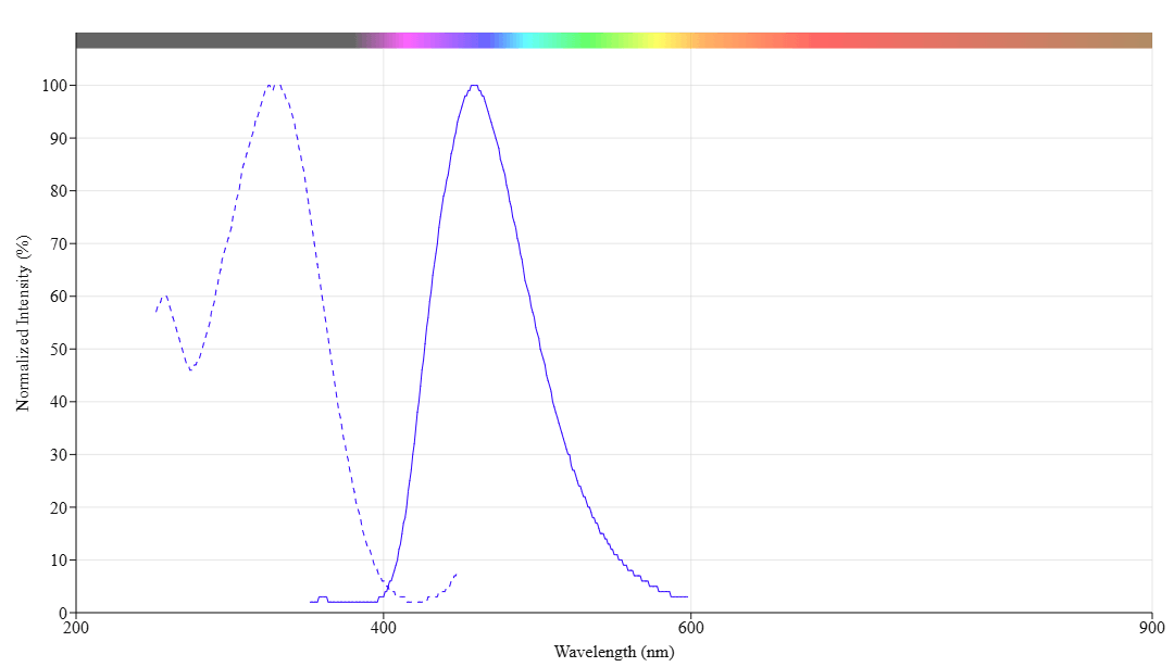
mFluor™ UVB 460 SE
mFluor™ UVB 460 SE is an amine-reactive dye optimally excited at 320 nm that provides a distinct blue emission channel for creating multiparameter flow cytometry panels with UV laser systems.
- Extended UV-to-Blue Emission: Excitation at ~320 nm with emission at ~460 nm creates an exceptionally large Stokes shift for reduced spectral crosstalk in multicolor panels
- Unique Spectral Niche: Fills a rare spectral window with minimal overlap with conventional fluorophores
- High Extinction Coefficient: Strong absorption (166,181 cm⁻¹M⁻¹) ensures efficient excitation and bright signal generation with standard UV laser lines
- Optimized for Abundant Markers: Small organic dye structure ideal for antibody conjugation, recommended for high-expression targets like CD4
| Catalog | Size | Price | Quantity |
|---|---|---|---|
| 70705 | 1 mg | Price |

Physical properties
| Molecular weight | 1093.38 |
| Solvent | DMSO |
Spectral properties
| Correction factor (280 nm) | 0.573 |
| Extinction coefficient (cm -1 M -1) | 16618 1 |
| Excitation (nm) | 325 |
| Emission (nm) | 459 |
Storage, safety and handling
| H-phrase | H303, H313, H333 |
| Hazard symbol | XN |
| Intended use | Research Use Only (RUO) |
| R-phrase | R20, R21, R22 |
| Storage | Freeze (< -15 °C); Minimize light exposure |
Contact us
| Telephone | |
| Fax | |
| sales@aatbio.com | |
| International | See distributors |
| Bulk request | Inquire |
| Custom size | Inquire |
| Technical Support | Contact us |
| Request quotation | Request |
| Purchase order | Send to sales@aatbio.com |
| Shipping | Standard overnight for United States, inquire for international |
Page updated on July 13, 2026