Products
Services
Resources
Selection Guides
About
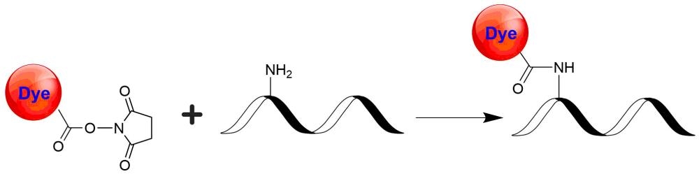
mFluor™ Blue 570 SE
mFluor™ Blue 570 dyes are an excellent alternative to RPE since they have the spectral properties equivalent to those of RPE conjugates. mFluor™ Blue 570 dyes are water-soluble, and the protein conjugates prepared with mFluor™ Blue 570 dyes are well excited at 488 nm to give red fluorescence (compatible with TRITC filter). mFluor™ Blue 570 dye and conjugates are excellent blue laser reagents for flow cytometry research. Compared to RPE, mFluor™ Blue 570 dyes are much more photostable, making them readily available for fluorescence imaging applications while it is very difficult to use RPE conjugates for fluorescence imaging applications due to the rapid photobleaching of RPE conjugates.
| Catalog | Size | Price | Quantity |
|---|---|---|---|
| 1160 | 1 mg | Price |

Physical properties
| Molecular weight | 1834.25 |
| Solvent | DMSO |
Spectral properties
| Absorbance (nm) | 553 |
| Correction factor (260 nm) | 0.228 |
| Correction factor (280 nm) | 0.179 |
| Extinction coefficient (cm -1 M -1) | 120000 1 |
| Excitation (nm) | 505 |
| Emission (nm) | 564 |
Storage, safety and handling
| H-phrase | H303, H313, H333 |
| Hazard symbol | XN |
| Intended use | Research Use Only (RUO) |
| R-phrase | R20, R21, R22 |
| Storage | Freeze (< -15 °C); Minimize light exposure |
| UNSPSC | 12171501 |
Contact us
| Telephone | |
| Fax | |
| sales@aatbio.com | |
| International | See distributors |
| Bulk request | Inquire |
| Custom size | Inquire |
| Technical Support | Contact us |
| Request quotation | Request |
| Purchase order | Send to sales@aatbio.com |
| Shipping | Standard overnight for United States, inquire for international |
Page updated on June 29, 2026