Phycoerythrin (PE)
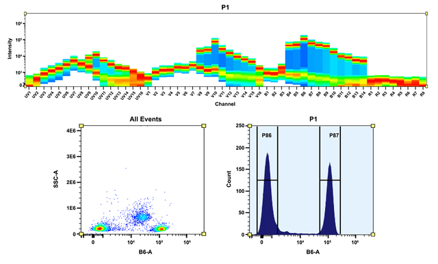
R-Phycoerythrin (PE) is isolated from red algae. Its primary absorption peak is at 565 nm with secondary peaks at 496 and 545 nm. The relative prominence of the secondary peaks varies significantly among R-PEs from different species. PE has three types of subunits: alpha (20,000 daltons), beta (20,000 daltons) and gamma (30,000 daltons). The molecular weight of intact PE has been found to be about 240,000 daltons. The alpha subunit of PE contains only the phycoerythrobilin (PEB) chromophore, while beta and gamma subunits contain both PEB and phycourobilin (PUB). Variability in the absorption spectra of PEs from various species reflects differences in the PEB/PUB ratio of the subunits. PE and closely related B-PE are the most intensely fluorescent phycobiliproteins, with quantum efficiencies probably in excess of 90%, and its orange fluorescence is readily visible by eye in any moderately concentrated solution.
Spectrum
Alternative formats
| Name | Form |
| Phycoerythrin (PE) | Purified |
| Phycoerythrin (PE) | Purified |
| Phycoerythrin (PE) | Purified |
| ReadiUse™ PE [R-Phycoerythrin] *Ammonium Sulfate-Free* | Ammonium Sulfate Free |
| ReadiUse™ PE [R-Phycoerythrin] *Ammonium Sulfate-Free* | Ammonium Sulfate Free |
| ReadiUse™ Preactivated PE Maleimide [Activated R-Phycoerythrin] | Maleimide |
| ReadiUse™ Preactivated PE Maleimide [Activated R-Phycoerythrin] | Maleimide |
Product family
| Name | Excitation (nm) | Emission (nm) | Extinction coefficient (cm -1 M -1) | Quantum yield | Correction Factor (280 nm) |
| ReadiUse™ PE [R-Phycoerythrin] *Ammonium Sulfate-Free* | 565 | 574 | 1960000 | 0.82 | 0.175 |
Citations
View all 9 citations: Citation Explorer
Tuning the In Vivo Photosynthetic Activity of Cyanobacteria’s Phycobilisome complex: A Spectroscopic Approach
Authors: Kalkar, Swapna and Ayerh, Jeffrey C and Herr, Daniel and Ignatova, Tetyana
Journal: (2025)
Authors: Kalkar, Swapna and Ayerh, Jeffrey C and Herr, Daniel and Ignatova, Tetyana
Journal: (2025)
Effectiveness evaluation of fluorescent compensation in multicolor flow cytometry: A quantitative study
Authors: Fan, Long and Gao, Chiyuan and Wang, Junbo and Huo, Xiaoye and Chen, Jian
Journal: Nanotechnology and Precision Engineering (2025)
Authors: Fan, Long and Gao, Chiyuan and Wang, Junbo and Huo, Xiaoye and Chen, Jian
Journal: Nanotechnology and Precision Engineering (2025)
In Vitro and In Vivo Metabolic Tagging and Modulation of Platelets
Authors: Baskaran, Dhyanesh and Liu, Yusheng and Zhou, Jiadiao and Wang, Yueji and Nguyen, Daniel and Wang, Hua
Journal: Materials Today Bio (2025): 101719
Authors: Baskaran, Dhyanesh and Liu, Yusheng and Zhou, Jiadiao and Wang, Yueji and Nguyen, Daniel and Wang, Hua
Journal: Materials Today Bio (2025): 101719
The Nedd4L ubiquitin ligase is activated by FCHO2-generated membrane curvature
Authors: Sakamoto, Yasuhisa and Uezu, Akiyoshi and Kikuchi, Koji and Kang, Jangmi and Fujii, Eiko and Moroishi, Toshiro and Suetsugu, Shiro and Nakanishi, Hiroyuki
Journal: The EMBO Journal (2024): 1--27
Authors: Sakamoto, Yasuhisa and Uezu, Akiyoshi and Kikuchi, Koji and Kang, Jangmi and Fujii, Eiko and Moroishi, Toshiro and Suetsugu, Shiro and Nakanishi, Hiroyuki
Journal: The EMBO Journal (2024): 1--27
Targeting pro-inflammatory T cells as a novel therapeutic approach to potentially resolve atherosclerosis in humans
Authors: Fan, Lin and Liu, Junwei and Hu, Wei and Chen, Zexin and Lan, Jie and Zhang, Tongtong and Zhang, Yang and Wu, Xianpeng and Zhong, Zhiwei and Zhang, Danyang and others,
Journal: Cell Research (2024): 1--21
Authors: Fan, Lin and Liu, Junwei and Hu, Wei and Chen, Zexin and Lan, Jie and Zhang, Tongtong and Zhang, Yang and Wu, Xianpeng and Zhong, Zhiwei and Zhang, Danyang and others,
Journal: Cell Research (2024): 1--21
References
View all 46 references: Citation Explorer
Chromophore attachment to phycobiliprotein beta-subunits: phycocyanobilin:cysteine-beta84 phycobiliprotein lyase activity of CpeS-like protein from Anabaena Sp. PCC7120
Authors: Zhao KH, Su P, Li J, Tu JM, Zhou M, Bubenzer C, Scheer H.
Journal: J Biol Chem (2006): 8573
Authors: Zhao KH, Su P, Li J, Tu JM, Zhou M, Bubenzer C, Scheer H.
Journal: J Biol Chem (2006): 8573
Excitation energy transfer from phycobiliprotein to chlorophyll d in intact cells of Acaryochloris marina studied by time- and wavelength-resolved fluorescence spectroscopy
Authors: Petrasek Z, Schmitt FJ, Theiss C, Huyer J, Chen M, Larkum A, Eichler HJ, Kemnitz K, Eckert HJ.
Journal: Photochem Photobiol Sci (2005): 1016
Authors: Petrasek Z, Schmitt FJ, Theiss C, Huyer J, Chen M, Larkum A, Eichler HJ, Kemnitz K, Eckert HJ.
Journal: Photochem Photobiol Sci (2005): 1016
Single-molecule spectroscopy selectively probes donor and acceptor chromophores in the phycobiliprotein allophycocyanin
Authors: Loos D, Cotlet M, De Schryver F, Habuchi S, Hofkens J.
Journal: Biophys J (2004): 2598
Authors: Loos D, Cotlet M, De Schryver F, Habuchi S, Hofkens J.
Journal: Biophys J (2004): 2598
Evaluation of Tolypothrix germplasm for phycobiliprotein content
Authors: Prasanna R, Prasanna BM, Mohammadi SA, Singh PK.
Journal: Folia Microbiol (Praha) (2003): 59
Authors: Prasanna R, Prasanna BM, Mohammadi SA, Singh PK.
Journal: Folia Microbiol (Praha) (2003): 59
Isolation and characterisation of phycobiliprotein rich mutant of cyanobacterium Synechocystis sp
Authors: Prasanna R, Dhar DW, Dominic TK, Tiwari ON, Singh PK.
Journal: Acta Biol Hung (2003): 113
Authors: Prasanna R, Dhar DW, Dominic TK, Tiwari ON, Singh PK.
Journal: Acta Biol Hung (2003): 113
Page updated on August 16, 2025


