logo
AAT Bioquest

Propidium Iodide

1 mg/mL aqueous solution
Propidium iodide is a DNA binding, red-fluorescent dye that can be used to stain dead cell populations in flow cytometry and fluorescence microscopy.

Propidium iodide (PI) belongs to a class of intercalating nucleic acid probes; this family of compounds includes other popular fluorophores such as acridine orange. Unlike acridine orange, however, PI contains two positive charges, which confers it high affinity for the negatively charged DNA in cells, but also prevents it from passing through the hydrophobic lipid bilayer. Because of this characteristic, propidium iodide is often used to quantify cell viability, by differentiating healthy cells, where it is excluded, from dead cells, whose compromised membranes allow staining.

Cell viability assessments in flow cytometry often make use of propidium iodide alongside acridine orange. These two probes are chosen because they are well excited by the ubiquitous 488 nm argon ion laser. In these assays, propidium iodide is used to identify dead cell populations while acridine orange, which is cell permeable, quantifies healthy cells.

Another flow cytometry application is the usage of propidium iodide in differentiating apoptosis and necrosis. In these applications, PI is paired with an Annexin V conjugate, such as Annexin V-FITC, wherein the former stains the necrotic cells, with compromised membranes, while the latter stains the apoptotic cells, by binding to translocated phosphatidylserine.

Staining principle

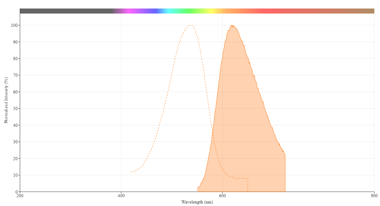
Propidium iodide binds DNA by intercalation, meaning it inserts itself between the planar geometries of nucleic acid base pairs. Unlike DAPI, which has a preference for A-T rich regions, PI shows no sequence specificity. In general, the stoichiometry is one dye molecule per 4-5 base pairs of DNA. Because of its double positive charge, propidium iodide has a high affinity for the negatively charged phosphate groups within DNA. After intercalation, the probe experiences a 20 to 30 fold increase in fluorescence, being excitable at 537 nm and emitting orange-red at 618 nm.

Excitation and emission

Propidium iodide is a fluorescent compound with an excitation peak at 537 nm and an emission peak at 618 nm. It is well excited by a 488 nm or 532 nm laser. It can be read using the PE/Texas Red filter set or the flow cytometry FL3 channel.

Solubility

Propidium iodide can be dissolved in several different solvents, both aqueous and organic, such as ethanol, DMSO, dimethyl formamide, PBS and water. The table below includes each solvent's respective solubility:

SolventSolubility
Ethanol0.2 mg / mL
DMSO2.5 mg / mL
Dimethyl formamide3.3 mg / mL
PBS2 mg / mL
Water1 mg / mL

Preparation of staining solution

Propidium iodide has a molecular weight of 668.39 daltons. The recommended staining concentration for flow cytometry is between 1 to 50 µg / mL depending on number of cells and experimental conditions. To prepare a 50 µg / mL solution from 1 mg / mL stock, dilute using PBS. Adding 50 µL of stock solution to 950 µL of PBS will yield a 1:20 dilution, resulting in 1 mL of 50 µg / mL propidium iodide working solution.

Flow cytometry staining protocol

This is an example for using propidium iodide in flow cytometry analysis. Optimization may be required for particular experimental conditions.

Prepare 7 mL of Fixing Solution
1X PBS
2% BSA
5 mM EDTA
0.1% NaN3

Prepare 500 µL of Sorting Buffer
1X PBS
0.1% Triton-X 100
2% BSA
5mM EDTA
40 µg / mL of propidium iodide
100 µg / mL RNase A

  1. Harvest and wash cells. For adherent cells, detach from growth surface using trypsin. For suspension cells, use centrifugation.
  2. Resuspend cells in 7 mL of Fixing Solution.
  3. Fix cells by adding 3 mL of 100% ethanol dropwise.
  4. Allow fixation for 30 minutes at 4 °C.
  5. Wash cells in PBS two times, spin at 2000 rpm in a centrifuge.
  6. Resuspend cells in 500 µL of Sorting Buffer.
  7. Incubate at 37 °C for 30 minutes.
  8. Analyze cells using flow cytometry with PE/Texas Red, FL2 or FL3 channels.

Spectrum

Product family

Citations

View all 14 citations: Citation Explorer
Chlorite and bromate alter the conjugative transfer of antibiotic resistance genes: Co-regulation of oxidative stress and energy supply
Authors: Cao, Jinrui and Xue, Bin and Yang, Shuran and Yang, Xiaobo and Zhang, Xi and Qiu, Zhigang and Shen, Zhiqiang and Wang, Jingfeng
Journal: Journal of Hazardous Materials (2024): 134257
Exploring Neuronal Differentiation Profiles in SH-SY5Y Cells through Magnetic Levitation Analysis
Authors: Kartal, Rumeysa Bilginer and Yildiz, Ahu Arslan
Journal: ACS omega (2024): 14955
Cylindrospermopsin enhances the conjugative transfer of plasmid-mediated multi-antibiotic resistance genes through glutathione biosynthesis inhibition
Authors: Yang, Shuran and Cao, Jinrui and Zhao, Chen and Zhang, Xi and Li, Chenyu and Wang, Shang and Yang, Xiaobo and Qiu, Zhigang and Li, Chao and Wang, Jingfeng and others,
Journal: Ecotoxicology and Environmental Safety (2024): 116288
Galectin-1 overexpression induces normal fibroblasts translate into cancer-associated fibroblasts and attenuates the sensitivity of anlotinib in lung cancer
Authors: Zhang, Lei and Chen, Wenbang and Li, Xiaojun and Wang, Gengming and Xing, Fubao and Zhu, Xiao
Journal: Cell Adhesion \& Migration (2024): 1--11
Microfluidic System for Cell Mixing and Particle Focusing Using Dean Flow Fractionation
Authors: Wiede, Alexander and Stranik, Ondrej and Tannert, Astrid and Neugebauer, Ute
Journal: (2023): 671--685

References

View all 50 references: Citation Explorer
Detection of gamma radiation processed onion during storage using propidium iodide based fluorescence microscopy.
Authors: Sharma, Tanmayee and Kavita, and Mishra, Bibhuti Bhusan and Variyar, Prasad Shekhar
Journal: Food chemistry (2023): 133928
Replacement of DAPI with propidium iodide could extend the utilisation of archival tissue samples for immunofluorescent techniques.
Authors: Cizkova, Katerina and Koubova, Katerina and Tauber, Zdenek
Journal: Histopathology (2022): 409-411
Corrigendum to: The DNA intercalators ethidium bromide and propidium iodide also bind to core histones.
Authors:
Journal: FEBS open bio (2022): 1087
Propidium iodide enabled live imaging of Pasteuria sp.-Pratylenchus zeae infection studies under fluorescence microscopy.
Authors: Perrine-Walker, Francine and Le, Khoa
Journal: Protoplasma (2021): 279-287
An Annexin V-FITC-Propidium Iodide-Based Method for Detecting Apoptosis in a Non-Small Cell Lung Cancer Cell Line.
Authors: Kumar, Robin and Saneja, Ankit and Panda, Amulya K
Journal: Methods in molecular biology (Clifton, N.J.) (2021): 213-223
Page updated on September 16, 2024

Ordering information

Price
FormAqueous solution
Aqueous solution
Powder
Concentration1 mg / mL
1 mg / mL
10 mM
Unit size
5 mL
10 mL
Catalog Number
Quantity
Add to cart

Additional ordering information

Telephone1-800-990-8053
Fax1-800-609-2943
Emailsales@aatbio.com
InternationalSee distributors
Bulk requestInquire
Custom sizeInquire
Technical SupportContact us
Purchase orderSend to sales@aatbio.com
ShippingStandard overnight for United States, inquire for international
Request quotation

Physical properties

Molecular weight

668.39

Solvent

Water

Spectral properties

Extinction coefficient (cm -1 M -1)

60001

Excitation (nm)

537

Emission (nm)

618

Quantum yield

0.21

Storage, safety and handling

Certificate of OriginDownload PDF
H-phraseH303, H313, H333
Hazard symbolXN
Intended useResearch Use Only (RUO)
R-phraseR20, R21, R22

Storage

Freeze (< -15 °C); Minimize light exposure

CAS

25535-16-4
Propidium iodide is a fluorescent compound with an excitation peak at 537 nm and an emission peak at 618 nm. Other spectra of interest include: 7-AAD (7-Aminoactinomycin D), DAPI (4,6-Diamidino-2-phenylindole), and Hoechst 33258. Propidium iodide belongs to the following categories: Cell Cycle Assays, Nucleus, Cell Viability Assays, Fluorescence Activated Cell Sorting (FACS), and Immunohistochemistry (IHC).
Effect of FSS on apoptosis and necrosis in tubular cells. Confluent monolayers of HK-2 cells were submitted to FSS 0 (static) or FSS 0.5 Pa (FSS 0.5) for 48h. A/ Cells were stained with Annexin-V and then immediately subjected to analysis of phosphatidylserine externalization (Annexin-V fluorescence, X-axis) and Propidium Iodide (PI) uptake (PI fluorescence, Y-axis) using flow cytometry. Living, early apoptotic or necrotic (primary or secondary) cells were distinguished by the criteria of Annexin-V-/PI-(bottom left quadrant), Annexin-V+/PI- (bottom right quadrant) and Annexin-V+/PI+ (upper right quadrant), respectively. B/ Proportions of early apoptosis and necrosis cells were quantified and results are expressed as a percentage of the total population of cells. Data represent mean ± SEM of 7 experiments. *HK-2 cells were assessed for apoptosis and necrosis using Cell Meter Annexin V Binding Apoptosis Assay Kit according to the manufacturer's instructions. Source: Shear Stress-Induced Alteration of Epithelial Organization in Human Renal Tubular Cells by Damien Maggiorani, et al., PLoS ONE, July 2015.
Gallery Image 3