Protonex™ Red 670-E. coli Conjugate
Product key features
The Protonex™ Red 670–E. coli Conjugate provides a ready-to-use, pH-sensitive fluorescent tool for monitoring phagocytosis and intracellular acidification processes in live cells.
- pH-activated fluorescence: Non-fluorescent at neutral pH and becomes strongly fluorescent in acidic compartments such as phagosomes and phagolysosomes.
- E. coli-based targeting: E. coli particles serve as biologically relevant substrates that mimic bacterial infection and are efficiently recognized by phagocytes.
- Multipurpose reagent: Suitable for incorporation into custom assay workflows for microscopy, flow cytometry, or plate-based readouts.
Product description
As a standalone reagent, it enables users to integrate phagocytic detection into their own custom assays. The Cy5-like excitation/emission properties of Protonex™ Red 670 make it compatible with a wide range of fluorescence imaging and detection systems. These conjugates can be used in combination with green fluorescent dyes like GFP, Calbryte™ 520, calcein AM, or FITC-labeled antibodies for multiplexed cell functional analysis. It is ideal for immunological research, drug discovery, and mechanistic studies of innate immune function, autophagy, or bacterial clearance.
Example protocol
AT A GLANCE
- Plate the cells.
- Treat cells with test compounds.
- Add Protonex Dye E. coli conjugates in medium.
- Incubate at 37°C for 60 minutes.
- Monitor fluorescence by microscope or fluorescence plate reader.
CELL PREPARATION
For guidelines on cell sample preparation, please visit:
https://www.aatbio.com/resources/guides/cell-sample-preparation.html
- Plate cells overnight in a growth medium at 20,000-50,000 cells/well/100 µL in a 96-well plate.
Note: For RAW 264.7 cells used in this assay, we recommend plating 50,000 cells per well in 100 µL of medium in a 96-well plate and incubating them overnight. It is important to optimize the cell density for each cell line individually.
Note: Higher background fluorescence levels may be seen with poly-D-lysine coated microplates.
SAMPLE EXPERIMENTAL PROTOCOL
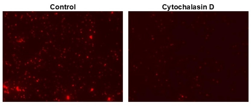
Add phagocytosis inhibitor (e.g., Cytochalasin D ) at the desired concentrations. You may need to add vehicle controls to untreated wells. (For example: 11X working solution can be prepared in PBS, and 10 µL can be added to each well.)
Note: The time and concentration of phagocytosis effectors varies with cell types.
- Add the suspension of E. coli conjugate to the cell culture microplate in a 1:10 dilution, or 10 μL of particles added to 100 μL of cell culture medium, and mix well.
- Place the cells at 37°C for 60 minutes to 3 hours.
- Wash the cells 2-3 times with HHBS Buffer (AAT Cat# 20011) or buffer of your choice.
- Add 100 µL HHBS Buffer to each well.
- Observe plate with a fluorescence microscope using the following filter set or read plate in a fluorescence plate reader with bottom read mode.
Spectrum

Product family
| Name | Excitation (nm) | Emission (nm) |
| Protonex™ Red 600-E. coli Conjugate | 576 | 597 |
References
Authors: Liu, Qian and Wang, Qinghua and Meng, Xiangchuan and Wang, Xiang and Zhang, Qingyang and Hu, Hai-Yu
Journal: ACS sensors (2025): 1072-1082
Authors: Liu, Songyun and Chen, Si and Lai, Barry and Antipova, Olga and Luo, Yanqi and Hall, Deborah J and Jin, Qiaoling and Maxey, Evan and Jacobs, Joshua J and Pourzal, Robin
Journal: Scientific reports (2025): 12467
Authors: Raneri, Simona and Gianoncelli, Alessandra and Bonanni, Valentina and Mirata, Serena and Scarfì, Sonia and Fornasini, Laura and Bersani, Danilo and Baroni, Debora and Picco, Cristiana and Gualtieri, Alessandro F
Journal: Environmental research (2024): 118878
Authors: Jiao, Mingxia and Li, Xiaoqi and Liu, Hui and Cai, Peng and Yang, Xiling and McHugh, Kevin J and Zheng, Bowen and Sun, Jiachen and Zhang, Peisen and Luo, Xiliang and Jing, Lihong
Journal: ACS nano (2024): 19038-19053
Authors: Hashimura, Hidenori and Kuwana, Satoshi and Nakagawa, Hibiki and Abe, Kenichi and Adachi, Tomoko and Sugita, Toyoko and Fujishiro, Shoko and Honda, Gen and Sawai, Satoshi
Journal: Cell structure and function (2024): 135-153