logo
AAT Bioquest

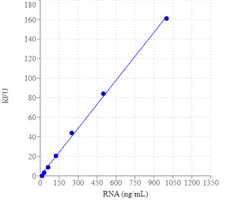
StrandBrite™ Green RNA Quantifying Reagent *200X DMSO Solution*

RNA dose response with StrandBrite™ Green in a solid black 96-well microplate and measured using a Gemini microplate reader (Molecular Devices).
Ordering information
Price
Catalog Number
Unit Size
Quantity
Add to cart
Additional ordering information
Telephone1-800-990-8053
Fax1-800-609-2943
Emailsales@aatbio.com
InternationalSee distributors
Bulk requestInquire
Custom sizeInquire
ShippingStandard overnight for United States, inquire for international
Request quotation
Physical properties
Molecular weightN/A
SolventDMSO
Spectral properties
Excitation (nm)509
Emission (nm)527
Storage, safety and handling
H-phraseH303, H313, H340
Hazard symbolT
Intended useResearch Use Only (RUO)
R-phraseR20, R21, R68
StorageFreeze (< -15 °C); Minimize light exposure
UNSPSC41116134

OverviewpdfSDSpdfProtocol


Molecular weight
N/A
Excitation (nm)
509
Emission (nm)
527
Detecting and quantitating small amounts of RNA is extremely important for a wide variety of molecular biology procedures such as measuring yields of in vitro transcribed RNA and measuring RNA concentrations before performing Northern blot analysis, S1 nuclease assays, RNase protection assays, cDNA library preparation, reverse transcription PCR, and differential display PCR. The most commonly used technique for measuring nucleic acid concentration is the determination of absorbance at 260 nm. The major disadvantage of the absorbance-based method is the interferences caused by proteins, free nucleotides and other UV absorbing compounds. The use of sensitive, fluorescent nucleic acid stains alleviates this interference problem. StrandBrite™ RNA quantifying reagent is an ultrasensitive fluorescent nucleic acid stain for quantitating RNA in solution. It is a positively charged fluorescent probe that binds to the hydrophobic pockets of RNA and forms a highly luminescent complex through the synergistic actions of stacking, hydrophobic forces, hydrogen bonding and electrostatic interactions. StrandBrite™ RNA quantifying reagent has extremely low inherent fluorescence that is significantly enhanced upon binding to RNAs, resulting in a great enhancement in its fluorescence. StrandBrite™ RNA quantifying reagent can detect as little as 5 ng/mL RNA with a fluorescence microplate reader or fluorometer.

Platform


Fluorescence microplate reader

Excitation490 nm
Emission525 nm
Cutoff515 nm
Recommended plateSolid black

Example protocol


PREPARATION OF STANDARD SOLUTION

For convenience, use the Serial Dilution Planner:
https://www.aatbio.com/tools/serial-dilution/17610


RNA Standard
Prepare a 100 ug/mL stock solution of RNA in DEPC treated water. Prepare 2 ug/mL in TE buffer from that stock and perform 1:2 dilutions to get 1000, 500, 250, 125, 62.5, 31.3, 15.6 and 0 ng/mL (Blank).

PREPARATION OF WORKING SOLUTION

StrandBrite™ Green working solution
Prepare an aqueous working solution of the StrandBrite™ Green by making a 200-fold dilution of the concentrated DMSO solution in TE (10 mM Tris-HCl, 1 mM EDTA, pH 7.5 in DEPC treated water). For example, add 50 μL StrandBrite™ Green to 10 mL TE buffer to prepare enough working solution to assay 100 samples in a 200 µL final volume. Protect the working solution from light by covering it with foil or placing it in the dark.
Note     We recommend preparing this solution in a plastic container rather than glass, as the dye may adsorb to glass surfaces.
Note     For the best results, this solution should be used within a few hours of its preparation.

SAMPLE EXPERIMENTAL PROTOCOL

The following protocol is an example for quantifying RNA with StrandBrite™ Green. Allow the StrandBrite™ Green to warm to room temperature before opening the vial.
Note     Always use clean disposable gloves while handling all materials to prevent RNase contamination.
Note     No data are available addressing the mutagenicity or toxicity of StrandBrite™ Green RNA stain. Because this reagent binds to nucleic acids, it should be treated as a potential mutagen and handled with appropriate care.

Table 1.Layout of RNA standards and test samples in a solid black 96-well microplate. (RS = RNA Standard; BL = Blank Control; TS = Test Samples)
BLBLTSTS
RS1RS1......
RS2RS2  
RS3RS3  
RS4RS4  
RS5RS5  
RS6RS6  
RS7RS7  
Table 2.Reagent composition for each well (Add the serially diluted RNA Standards from 15.6 to 1000 ng/mL into wells from RS1 to RS7 in duplicate)
RNA StandardBlank ControlTest Sample
Serial Dilutions (100 µL)TE: 100 µL100 µL

Run RNA assay
  1. Add 100 μL of StrandBrite™ Green working solution to each well of the RNA standard, blank control, and test samples to make the total RNA assay volume of 200 µL/well.
    Note     For a 384-well plate, add 25 μL sample and 25 μL of StrandBrite™ Green working solution per well.
  2. Incubate the reaction at room temperature for 5 to 10 minutes, protected from light.
  3. Monitor the fluorescence increase with a spectrofluorometer at Ex/Em = 490/525 nm (cutoff at 515 nm).
    Note      To minimize photobleaching effects, keep the time for fluorescence measurement constant for all samples.
  4. The fluorescence in blank wells (with the assay buffer only) is used as a control, and is subtracted from the values for those cuvettes with RNA standard or test samples. The RNA concentration of the sample is determined according to the RNA standard curve. 

Spectrum


Open in Advanced Spectrum Viewer
spectrum

Spectral properties

Excitation (nm)509
Emission (nm)527

Images


Citations


View all 3 citations: Citation Explorer
The nucleolar shell provides anchoring sites for DNA untwisting
Authors: Fukute, Jumpei and Maki, Koichiro and Adachi, Taiji
Journal: Communications Biology (2024): 83
Fine-tuning of highly bright benzo [c, d] indole-oxazolopyridine cyanine dye for nucleolar RNA imaging in living cells
Authors: Togashi, Nao and Nagaoka, Masaaki and Higuchi, Kei and Yoshino, Yukina and Wu, Yawen and Sato, Yusuke and Nishizawa, Seiichi
Journal: Talanta Open (2024): 100308
Anticancer Activity of Reconstituted Ribonuclease S-Decorated Artificial Viral Capsid
Authors: Liang, Yingbing and Furukawa, Hiroto and Sakamoto, Kentarou and Inaba, Hiroshi and Matsuura, Kazunori
Journal: ChemBioChem (2022): e202200220

References


View all 31 references: Citation Explorer
Inhibitors of Streptococcus pneumoniae surface endonuclease EndA discovered by high-throughput screening using a PicoGreen fluorescence assay
Authors: Peterson EJ, Kireev D, Moon AF, Midon M, Janzen WP, Pingoud A, Pedersen LC, Singleton SF.
Journal: J Biomol Screen (2013): 247
Validation of a PicoGreen-based DNA quantification integrated in an RNA extraction method for two-dimensional and three-dimensional cell cultures
Authors: Chen Y, Sonnaert M, Roberts SJ, Luyten FP, Schrooten J.
Journal: Tissue Eng Part C Methods (2012): 444
Characterization of PicoGreen interaction with dsDNA and the origin of its fluorescence enhancement upon binding
Authors: Dragan AI, Casas-Finet JR, Bishop ES, Strouse RJ, Schenerman MA, Geddes CD.
Journal: Biophys J (2010): 3010
Comparison of SYBR Green I-, PicoGreen-, and [3H]-hypoxanthine-based assays for in vitro antimalarial screening of plants from Nigerian ethnomedicine
Authors: Abiodun OO, Gbotosho GO, Ajaiyeoba EO, Happi CT, Hofer S, Wittlin S, Sowunmi A, Brun R, Oduola AM.
Journal: Parasitol Res (2010): 933
Metal-enhanced PicoGreen fluorescence: application to fast and ultra-sensitive pg/ml DNA quantitation
Authors: Dragan AI, Bishop ES, Casas-Finet JR, Strouse RJ, Schenerman MA, Geddes CD.
Journal: J Immunol Methods (2010): 95
Quantification of dsDNA using the Hitachi F-7000 Fluorescence Spectrophotometer and PicoGreen dye
Authors: Moreno LA, Cox KL.
Journal: J Vis Exp. (2010)
Factors affecting quantification of total DNA by UV spectroscopy and PicoGreen fluorescence
Authors: Holden MJ, Haynes RJ, Rabb SA, Satija N, Yang K, Blasic JR, Jr.
Journal: J Agric Food Chem (2009): 7221
Development and characterization of a novel host cell DNA assay using ultra-sensitive fluorescent nucleic acid stain "PicoGreen"
Authors: Ikeda Y, Iwakiri S, Yoshimori T.
Journal: J Pharm Biomed Anal (2009): 997
Enhanced DNA dynamics due to cationic reagents, topological states of dsDNA and high mobility group box 1 as probed by PicoGreen
Authors: Noothi SK, Kombrabail M, Kundu TK, Krishnamoorthy G, Rao BJ.
Journal: FEBS J (2009): 541
Label-free DNA sequence detection with enhanced sensitivity and selectivity using cationic conjugated polymers and PicoGreen
Authors: Ren X, Xu QH.
Journal: Langmuir (2009): 43