Cell Adhesion
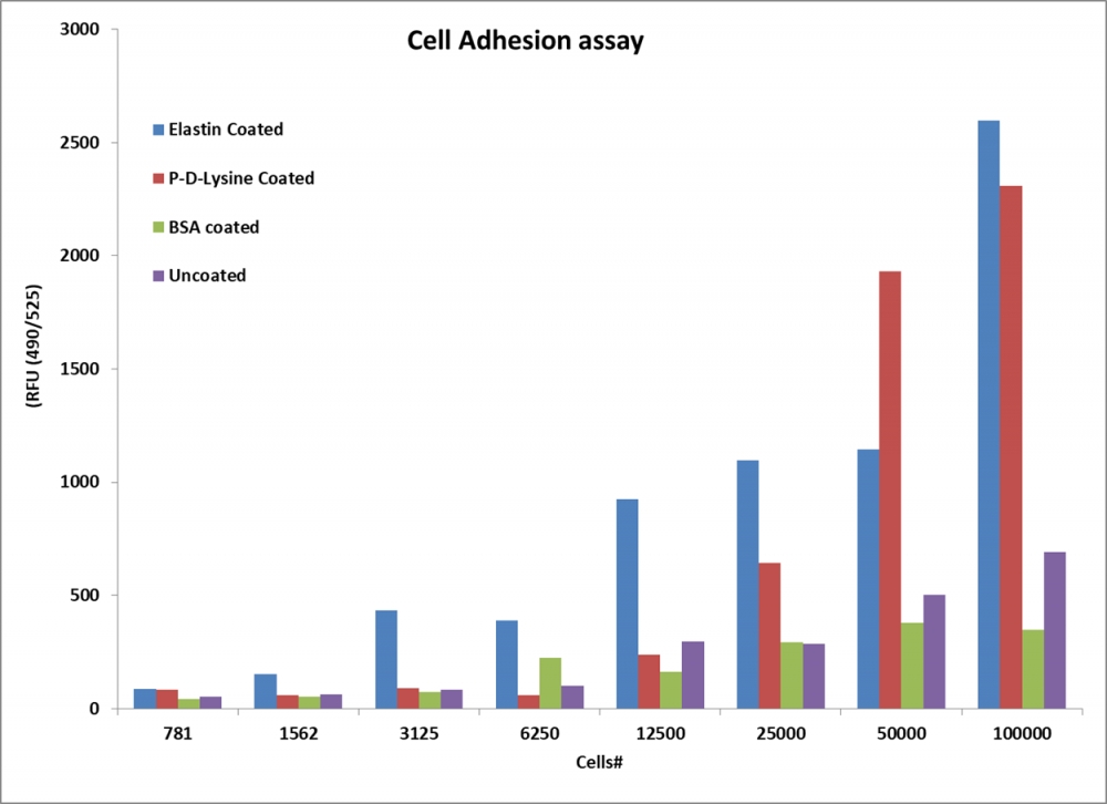
Cell adhesion measured with Cell Meter™ Cell Adhesion Assay Kit using a fluorescence microplate reader. Jurkat cells at different confluences or confluency levels were incubated in wells coated with different materials, and then stained with Calcein Ultragreen AM at 37°C for 30 mins. The signal was monitored at Ex/Em = 490/525 nm.
Cell adhesion assays help researchers and clinical investigators alike mimic specific cellular microenvironments in bioengineering prospects, biomaterial development and biopharmaceutical experimentation. The procedures are normally geared towards mammalian cell lines.
Table of Contents
- Cell Adhesion Assays
- Techniques in Adhesion Assays
- 2.1 Polyacrylamide-Traction Force Microscopy (PA-TFM)
- 2.2 Microfabricated Elastomeric Post Array (mPADs)
- 2.3 Three-Dimensional Traction Force Quantification (3D-TFM)
- 2.4 Wash Assay
- 2.5 Flow Adhesion
- 2.6 Resonance Frequency
- Product Ordering Information
- References
Cell Adhesion Assays
Cell adhesion requirements are dependent on the application of the cellular product, so various techniques have been employed for appropriate fields, like advancements in tissue engineering, biochemical or toxicological studies, and to better understand properties of some cancers. Generally, cell adhesion assays can exist in two distinct experimental categories, divided between static and flow experimental setups.
Static assays can be used for multiple types of cells, including epithelial and fibroblasts, or for the purpose of scaffolds in tissue generation. Static assays are relatively easy to perform, though time consuming, and are ideal for studying the cell interaction between ECM proteins, like fibronectin, linked to cell proliferation and biosynthesis.
Flow assays are used to better stimulate cell adhesion that occurs in blood, or even lymph vessels and heart valves, under shear stress using microfluidic chambers. These assays are more dynamic in nature, and provide in-depth physiological data with microscopic analysis; however they are not well suited for high-throughput experiments. Flow assays can be used to study leukocytes with endothelial cells or purified ligands, though they may also be used to study bacterial adhesion as well.
| Datasets: |
Techniques in Adhesion Assays
Adhesion and attachment experiments can be grouped into assays that study single cells or cell populations as a whole. Static and flow assays also help provide better understanding of specific adhesion kinetics. Some methods of commonly used cell adhesion assays are listed below.
Polyacrylamide-Traction Force Microscopy (PA-TFM)
The PA-TFM method is a 2D technique that measures substrate deformations induced by individual cells that can be fine-tuned mechanically as well as chemically. First, the PA substrate is functionalized with adhesive ligands, and then it is embedded with fluorescent beads before cells are cultured on the surface. Deformities in the gel formed from the cell culture dislocate the fluorescent beads, which displacement can be imaged and quantified via TFM. Benefits to this method are that PA is inexpensive, and 2D capabilities allow for basic understanding of cell migratory properties. The adaptability of PA under different conditions suggests that this assay also has benefits in mechanobiology research.
| Basis Of Differentiation | Adherent cell culture | suspension cell culture |
| Definition | Refers to cells that are grown in a single layer attached to a solid surface, such as a petri dish or flask supplemented with a culture medium | Refers to cells that are grown free-floating or suspended in a liquid medium either as single cells or as free-floating clusters |
| Suitability | Suitable for most cell types, including primary cultures | Suitable only for non-adhesive cell lines and for cells adapted to suspension culture |
| Special requirement | Requires a tissue culture treated vessel | Does not require a tissue culture treated vessel |
| Agitation | Does not require agitation during growth | Requires agitation during growth for adequate gas exchange |
| Anchorage dependency | Is anchorage dependent | Is anchorage independent |
| Trypsinization | Is present | Is absent |
| Dissociation | Cells can be dissociated enzymatically or mechanically | Cells do not require enzymatic or mechanical dissociation |
| Growth limitations | Growth is limited by surface area, which may limit product yields | Growth is limited by cell density in the medium |
| Passaging requirement | Requires periodic passaging, but allows easy visual inspection under inverted microscope | Easier to passage, but requires daily cell counts and viability determination in order to follow growth patterns |
| Yield | Low-yielding | High-yielding |
| Applications | Used for harvesting products continuously, cytology, and many different research applications | Used for batch harvesting, bulk protein production, and a wide range of research applications |
Microfabricated Elastomeric Post Array (mPADs)
mPADs, also known as micropatterning, is a 2D assay where a silicone or PDMS substrate with an organization of microposts (micropillars) is made using soft lithography. The micropillars are meticulously functionalized with an ECM protein before cells are passively applied, then deflective resultant forces are calculated to provide discrete, reliable, quantification. mPADs are highly sensitive, and can be used both for single cells or a population of cells, but require sophisticated machinery and optimized methods. Micropatterning better demonstrates the connection between biochemical and mechanical happenings in a cellular setting, with uses in tissue engineering and regenerative medicine.
Three-Dimensional Traction Force Quantification (3D-TFM)
Many cell types, like cancer cells, stem cells and fibroblasts behave differently in 3D cultures, due to having advanced cytoskeletal roles. Physiologically, cancer cells have exhibited notably dissimilar behaviors in 3D versus 2D in areas of gene expression, drug resistance, and metastatic reaction, signifying the importance of 3D assays that quantify spatial as well as temporal values. Such matrices can be composed of a number of different gels, like agarose, collagen, hyaluronic acid, fibrin, or PEG, and are visualized in a similar manner to 2D-TFM fluorescence. Though still a relatively new tool in the field of biomechanics, 3D-TFM has shown possibilities in characterizing previously not-well understood cell populations, differentiation, and migratory properties in a more biological setting.
| Application Notes: |
Wash Assay

Effects of Hpgd on the adhesion and angiogenesis of hPAECs. (A) The microscope results showed that OE-Hpgd reduced the adhesion of hPAECs. (B) Statistical histogram of the adherent cells. (C) The microscope results showed that OE-Hpgd reduced the angiogenesis capability. (D) Statistical bar chart of the number of tubules. (**P<0.01). Adhesion experiments of ECs were performed according to the instructions of the cell adhesion kit. Source: Hpgd affects the progression of hypoxic pulmonary hypertension by regulating vascular remodeling by Meng He, Kelong Tao, Min Xiang & Jian Sun. BMC Pulmonary Medicine volume 23, Article number: 116. April 2023.
Flow Adhesion
Microfluidic assays counter static measure by using low fluid shear flow techniques to aid with cell attachment, mimicking bodily blood flow. Cells are subject to biologically similar hemodynamic forces that can visualize cell adherence, spreading, and tracking in a physiological system. Microfluidic channel approaches have advanced to applications on tissue- and organ-on-a-chip studies, where microscopic channels are internally coated with cellular layers to replicate human vascularity. Cell adhesion can then be monitored inside and outside the channels, visualized, and quantitated for functional properties.
Resonance Frequency
In the static resonance frequency technique, a quartz crystal is sandwiched between two metal electrodes, and then coated with a target ECM to create a piezoelectric acoustic wave resonator. After cells are mounted onto the biosensor, it is placed in a chamber and changes in resonant frequency can be detected and tracked upon cell adhesion and migration in real time. Though results are highly accurate, experimentation can be costly and require field expertise in operation.
Product Ordering Information
Table 1. Cell Adhesion Assay Kits
| Cat# ▲ ▼ | Product Name ▲ ▼ | Unit Size ▲ ▼ |
| 23010 | Cell Meter™ Cell Adhesion Assay Kit | 100 Tests |
References
- Khalili, Amelia, and Mohd Ahmad. “A Review of Cell Adhesion Studies for Biomedical and Biological Applications.” International Journal of Molecular Sciences, vol. 16, no. 8, 2015, pp. 18149-18184., https://doi.org/10.3390/ijms160818149.
- Kucik D.F., Wu C. (2005) Cell-Adhesion Assays. In: Guan JL. (eds) Cell Migration. Methods in Molecular Biology™, vol 294. Humana Press. https://doi.org/10.1385/1-59259-860-9:043
- Zhou, Dennis W, and Andrés J García. “Measurement systems for cell adhesive forces.” Journal of biomechanical engineering vol. 137,2 (2015): 020908. doi:10.1115/1.4029210
- Tan, J. L., et al. “Cells Lying on a Bed of Microneedles: An Approach to Isolate Mechanical Force.” Proceedings of the National Academy of Sciences, vol. 100, no. 4, 2003, pp. 1484-1489., https://doi.org/ 10.1073/pnas.0235407100.
- Wegener, Joachim, Andreas Janshoff, and H-J. Galla. "Cell adhesion monitoring using a quartz crystal microbalance: comparative analysis of different mammalian cell lines." European biophysics journal 28.1 (1998): 26-37.