Products
Services
Resources
Selection Guides
About
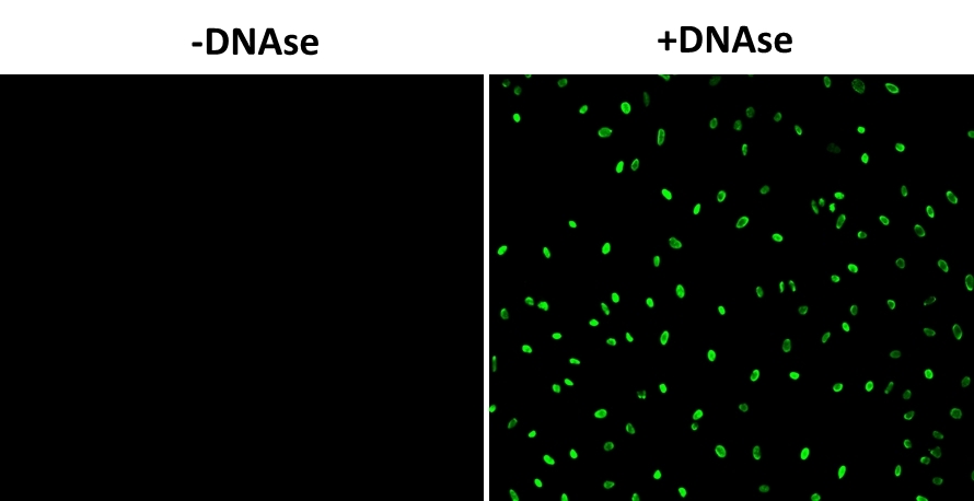
Cell Meter™ Fixed Cell and Tissue TUNEL Apoptosis Assay Kit
Green Fluorescence
Cell Meter™ Fixed Cell and Tissue TUNEL Apoptosis Assay Kit provides a robust tool for conveniently detecting DNA fragmentation caused by apoptosis. The assay is a non-radioactive, simple, accurate and rapid method for monitoring apoptosis in. fixed cells and tissues via imaging DNA fragmentation. The TUNEL assay uses terminal deoxynucleotidyl transferase (TdT) to catalyze the incorporation of fluorescein-12-dUTP at the 3’-hydroxyl ends of the fragmented DNA. The fluorescein-labeled DNA is analyzed by fluorescence microscopy or flow cytometry (excitation at 488 nm with 530/30 nm emission filter). The kit can be used to detect apoptosis in fixed cells and formalin-fixed, paraffin-embedded tissue sections.
| Catalog | Size | Price | Quantity |
|---|---|---|---|
| 22851 | 25 Tests | Price |

Spectral properties
| Absorbance (nm) | 487 |
| Correction factor (260 nm) | 0.32 |
| Correction factor (280 nm) | 0.35 |
| Extinction coefficient (cm -1 M -1) | 80000 1 |
| Excitation (nm) | 498 |
| Emission (nm) | 517 |
| Quantum yield | 0.7900 1 , 0.952 |
Storage, safety and handling
| H-phrase | H303, H313, H333 |
| Hazard symbol | XN |
| Intended use | Research Use Only (RUO) |
| R-phrase | R20, R21, R22 |
| UNSPSC | 12171501 |
Instrument settings
| Flow cytometer | |
| Excitation | 488 nm laser |
| Emission | 530/30 nm filter |
| Instrument specification(s) | FITC channel |
| Fluorescence microscope | |
| Excitation | FITC filter set |
| Emission | FITC filter set |
| Recommended plate | Black wall/clear bottom |
Contact us
| Telephone | |
| Fax | |
| sales@aatbio.com | |
| International | See distributors |
| Bulk request | Inquire |
| Custom size | Inquire |
| Technical Support | Contact us |
| Request quotation | Request |
| Purchase order | Send to sales@aatbio.com |
| Shipping | Standard overnight for United States, inquire for international |
Page updated on October 8, 2024