Cell Meter™ Fixed Cell and Tissue TUNEL Apoptosis Assay Kit *Red Fluorescence*
Ordering information
Price | |
Catalog Number | |
Unit Size | |
Quantity |
Additional ordering information
Telephone | 1-800-990-8053 |
Fax | 1-800-609-2943 |
sales@aatbio.com | |
International | See distributors |
Bulk request | Inquire |
Custom size | Inquire |
Shipping | Standard overnight for United States, inquire for international |
Spectral properties
Correction Factor (260 nm) | 0.27 |
Correction Factor (280 nm) | 0.34 |
Extinction coefficient (cm -1 M -1) | 100000 |
Excitation (nm) | 544 |
Emission (nm) | 570 |
Storage, safety and handling
H-phrase | H303, H313, H333 |
Hazard symbol | XN |
Intended use | Research Use Only (RUO) |
R-phrase | R20, R21, R22 |
UNSPSC | 12171501 |
Alternative formats
Related products
Overview | ![]() ![]() |
See also: DNA Fragmentation, TUNEL Assays
Correction Factor (260 nm) 0.27 | Correction Factor (280 nm) 0.34 | Extinction coefficient (cm -1 M -1) 100000 | Excitation (nm) 544 | Emission (nm) 570 |
Cell Meter™ TUNEL apoptosis assay kit provides a robust tool for conveniently detecting apoptosis caused by DNA fragmentation. The assay is non-radioactive and rapid. The TUNEL assay uses the terminal deoxynucleotidyl transferase (TdT) to catalyze the incorporation of TMR-dUTP at the free 3’-hydroxyl ends of the fragmented DNAs. The resulted TMR-labeled DNAs are analyzed by fluorescence microscopy (Cy3 or TRITC filter set). Its red emission can be conveniently multiplexed with GFP labelled targets. Direct incorporation of fluorescent TMR-labeled nucleotides significantly reduces the number of test steps. The kit is optimized to detect apoptosis in fixed cells and formalin-fixed, paraffin-embedded tissue sections.
Platform
Flow cytometer
Excitation | 488 nm laser |
Emission | 575/26 nm filter |
Instrument specification(s) | PE channel |
Fluorescence microscope
Excitation | Cy3 filter set |
Emission | Cy3 filter set |
Recommended plate | Black wall/clear bottom |
Components
Example protocol
AT A GLANCE
Protocol summary
- Treat samples as desired
- Fix cells with 4% formaldehyde solution for 30 minutes on ice
- Permeabilize cells with 70% ice-cold ethanol for 60 minutes on ice
- Add TdT staining solution to samples and incubate for 60 minutes at 37 °C
- Monitor the fluorescence intensity using fluorescence microscopy with Cy3 filter set
PREPARATION OF WORKING SOLUTION
TdT staining solution
For one test, Mix the following to make total volume of 51 µL;45 µL TdT Reaction Buffer (Component D)
5 µL CoCl2 (Component C)
0.5 µL TF5-dUTP (Component B)
0.5 µL TdT enzyme (Component A).
Note TdT staining solution should be used promptly.
SAMPLE EXPERIMENTAL PROTOCOL
The following protocol can be used as a guideline and should be optimized according to the needs.
Deparaffinization and rehydration protocol
- Treat your samples as desired.
- Wash the cells with buffer of your choice such as PBS containing Ca+2 and Mg+2.
- Fix the cells by adding 100 µL of 4% paraformaldehyde in PBS and incubate the samples for 30 minutes on ice.
- Remove fixation solution and wash cells with PBS.
- Add 100 µL of 70% of ice cold ethanol to cells and incubate the samples for 60 minutes on ice.
Note Cells can be stored at -20 °C at this step for several days before use. - Remove alcohol and wash cells with PBS.
Note For a positive control, incubate fixed cells with 2-5 µg/mL of DNAse in PBS containing Ca+2 and Mg+2 for 60 minutes at 37 °C. Remove the DNAse and wash cells thoroughly and continue with the rest of the protocol. - Add 50 µL of TdT staining solution to the cells and incubate for 60 to 120 minutes at 37 °C.
- Remove TdT working solution and wash cells with PBS.
- Resuspend the cells in PBS and monitor the fluorescence intensity with flow cytometer using 575/26 nm filter (PE channel) or fluorescence microscope with Cy3 filter set.
Protocol for tissue staining
The following protocol can be used as a guideline and should be optimized according to the needs.Deparaffinization and rehydration protocol
- Deparaffinize tissue sections (attached to the microscopic slides) by immersing slides in fresh xylene in a Coplin jar for 5 minutes at room temperature. Repeat one more time. (Total 2 washes)
- Wash the samples by immersing the slides in 100% ethanol for 5 minutes at room temperature in a Coplin jar.
- Rehydrate the samples by immersing the slides through various concentrations of alcohol subsequently (100, 95, 85, 70, 50%) for 5 minutes each at room temperature.
- Wash the samples by immersing the slides in 0.85% NaCl for 5 minutes at room temperature.
- Wash the samples by immersing the slides in PBS for 5 minutes at room temperature. Repeat one more wash. (Total 2 washes)
- Fix the tissue sections by immersing slides in 4% paraformaldehyde solution in PBS for 15-20 minutes at room temperature.
- Wash the samples by immersing the slides in PBS for 5 minutes at room temperature. Repeat one more wash. (Total 2 washes)
- Remove the liquid and place the slides on a flat surface. Treat tissue sections with 100 µL of 20 µg/mL Proteinase K solution. Add enough to cover the entire tissue surface. Incubate slides for 10 minutes at room temperature.
- Wash the samples by immersing the slides in PBS for 5 minutes at room temperature.
- Fix the tissue sections by immersing slides in 4% paraformaldehyde solution in PBS for 15-20 minutes at room temperature.
- Wash the samples by immersing the slides in PBS for 5 minutes at room temperature. Repeat one more wash. (Total 2 washes)
- Optional: For a positive control, incubate fixed samples with 2-5 µg/mL of DNAse in PBS containing Ca+2 and Mg+2 for 60 minutes at 37 °C. Remove the DNAse and wash cells thoroughly with PBS and continue with the rest of the protocol.
- Add 50 µL of TdT staining solution to the samples and incubate for 60 to 120 minutes at 37 °C.
- Remove TdT working solution and wash samples with PBS.
- Add mounting medium with DAPI (AAT Bioquest Cat# 20005) and monitor the fluorescence intensity fluorescence microscope with Cy3 filter set.
Spectrum
Open in Advanced Spectrum Viewer


Spectral properties
Correction Factor (260 nm) | 0.27 |
Correction Factor (280 nm) | 0.34 |
Extinction coefficient (cm -1 M -1) | 100000 |
Excitation (nm) | 544 |
Emission (nm) | 570 |
Product Family
Name | Excitation (nm) | Emission (nm) | Extinction coefficient (cm -1 M -1) | Correction Factor (260 nm) | Correction Factor (280 nm) |
Cell Meter™ Fixed Cell and Tissue TUNEL Apoptosis Assay Kit *Blue Fluorescence* | 411 | 472 | - | 0.14 | 0.12 |
Cell Meter™ Fixed Cell and Tissue TUNEL Apoptosis Assay Kit *Green Fluorescence* | 498 | 517 | 800001 | 0.32 | 0.35 |
Cell Meter™ Fixed Cell and Tissue TUNEL Apoptosis Assay Kit *Deep Red Fluorescence* | 649 | 664 | 250000 | - | 0.027 |
Images

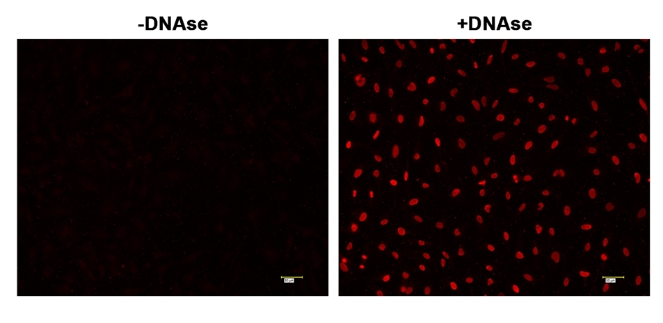
Figure 1. Fluorescence images of TUNEL assay with HeLa cells.
HeLa cells were fixed and treated with or without DNAse for 60 mins at 37 °C. The cells were then stained with Cell Meter™ Fixed Cell and Tissue TUNEL Apoptosis Assay Kit. DNA strand breaks showed intense fluorescent staining in DNAse treated cells. The signal was acquired with fluorescence microscope using a Cy3 filter set.
Citations
View all 8 citations: Citation Explorer
Antiapoptotic and antioxidant effects of melatonin on cat vitrified oocytes
Authors: Colombo, M and Mascaro, A and Pecile, A and Fusi, J and Luvoni, GC and others,
Journal: REPRODUCTION IN DOMESTIC ANIMALS (2023): 192--192
Authors: Colombo, M and Mascaro, A and Pecile, A and Fusi, J and Luvoni, GC and others,
Journal: REPRODUCTION IN DOMESTIC ANIMALS (2023): 192--192
Effects of boric acid on invasion, migration, proliferation, apoptosis and miRNAs in medullary thyroid cancer cells
Authors: Y{\i}ld{\i}r{\i}m, Onurcan and Se{\c{c}}me, M{\"u}cahit and Dodurga, Yavuz and Mete, G{\"u}l{\c{c}}in Abban and Fenkci, Semin Melahat
Journal: (2023)
Authors: Y{\i}ld{\i}r{\i}m, Onurcan and Se{\c{c}}me, M{\"u}cahit and Dodurga, Yavuz and Mete, G{\"u}l{\c{c}}in Abban and Fenkci, Semin Melahat
Journal: (2023)
Pterostilbene-Isothiocyanate Inhibits Proliferation of Human MG-63 Osteosarcoma Cells via Abrogating $\beta$-Catenin/TCF-4 Interaction─ A Mechanistic Insight
Authors: Kumar, Viney and Haldar, Swati and Ghosh, Souvik and Saini, Saakshi and Dhankhar, Poonam and Roy, Partha
Journal: ACS Omega (2023)
Authors: Kumar, Viney and Haldar, Swati and Ghosh, Souvik and Saini, Saakshi and Dhankhar, Poonam and Roy, Partha
Journal: ACS Omega (2023)
Inhibition of lung microbiota-derived proapoptotic peptides ameliorates acute exacerbation of pulmonary fibrosis
Authors: D’Alessandro-Gabazza, Corina N and Yasuma, Taro and Kobayashi, Tetsu and Toda, Masaaki and Abdel-Hamid, Ahmed M and Fujimoto, Hajime and Hataji, Osamu and Nakahara, Hiroki and Takeshita, Atsuro and Nishihama, Kota and others,
Journal: Nature communications (2022): 1--23
Authors: D’Alessandro-Gabazza, Corina N and Yasuma, Taro and Kobayashi, Tetsu and Toda, Masaaki and Abdel-Hamid, Ahmed M and Fujimoto, Hajime and Hataji, Osamu and Nakahara, Hiroki and Takeshita, Atsuro and Nishihama, Kota and others,
Journal: Nature communications (2022): 1--23
Protection of propofol on liver ischemia reperfusion injury by regulating Cyp2b10/Cyp3a25 pathway
Authors: Wu, Jinli and Yu, Chao and Zeng, Xianggang and Xu, Yini and Sun, Chengyi
Journal: Tissue and Cell (2022): 101891
Authors: Wu, Jinli and Yu, Chao and Zeng, Xianggang and Xu, Yini and Sun, Chengyi
Journal: Tissue and Cell (2022): 101891
Reduction of Amyloid Burden by Proliferated Homeostatic Microglia in Toxoplasma gondii-Infected Alzheimer's Disease Model Mice
Authors: Shin, Ji-Hun and Hwang, Young Sang and Jung, Bong-Kwang and Seo, Seung-Hwan and Ham, Do-Won and Shin, Eun-Hee
Journal: International journal of molecular sciences (2021): 2764
Authors: Shin, Ji-Hun and Hwang, Young Sang and Jung, Bong-Kwang and Seo, Seung-Hwan and Ham, Do-Won and Shin, Eun-Hee
Journal: International journal of molecular sciences (2021): 2764
Function-adaptive clustered nanoparticles reverse Streptococcus mutans dental biofilm and maintain microbiota balance
Authors: Ostadhossein, Fatemeh and Moitra, Parikshit and Altun, Esra and Dutta, Debapriya and Sar, Dinabandhu and Tripathi, Indu and Hsiao, Shih-Hsuan and Kravchuk, Valeriya and Nie, Shuming and Pan, Dipanjan
Journal: Communications Biology (2021): 1--16
Authors: Ostadhossein, Fatemeh and Moitra, Parikshit and Altun, Esra and Dutta, Debapriya and Sar, Dinabandhu and Tripathi, Indu and Hsiao, Shih-Hsuan and Kravchuk, Valeriya and Nie, Shuming and Pan, Dipanjan
Journal: Communications Biology (2021): 1--16
Exosomes Derived from M2 Macrophages Exert a Therapeutic Effect via Inhibition of the PI3K/AKT/mTOR Pathway in Rats with Knee Osteoarthritic
Authors: Da-Wa, Zha Xi and Jun, Ma and Chao-Zheng, Liu and Sen-Lin, Yang and Chuan, Lu and De-Chun, Li and Zu-Nan, Dong and Hong-Tao, Zhao and Shu-Qing, Wei and Xian-Wei, Pei and others,
Journal: BioMed Research International (2021)
Authors: Da-Wa, Zha Xi and Jun, Ma and Chao-Zheng, Liu and Sen-Lin, Yang and Chuan, Lu and De-Chun, Li and Zu-Nan, Dong and Hong-Tao, Zhao and Shu-Qing, Wei and Xian-Wei, Pei and others,
Journal: BioMed Research International (2021)
References
View all 21 references: Citation Explorer
Vitex Rotundifolia Fractions Induced Apoptosis in Human Breast Cancer T-47D Cell Line via Activation of Extrinsic and Intrinsic Pathway.
Authors: Chaudhry, Gul-E-Saba and Jan, Rehmat and Naveed Zafar, Muhammad and Mohammad, Habsah and Muhammad, Tengku Sifzizul Tengku
Journal: Asian Pacific journal of cancer prevention : APJCP (2019): 3555-3562
Authors: Chaudhry, Gul-E-Saba and Jan, Rehmat and Naveed Zafar, Muhammad and Mohammad, Habsah and Muhammad, Tengku Sifzizul Tengku
Journal: Asian Pacific journal of cancer prevention : APJCP (2019): 3555-3562
A mechanism for semaphorin-induced apoptosis: DNA damage of endothelial and myogenic cells in primary cultures from skeletal muscle.
Authors: Hei Yuan, Haynes Shek and Katyal, Sachin and Anderson, Judy E
Journal: Oncotarget (2018): 22618-22630
Authors: Hei Yuan, Haynes Shek and Katyal, Sachin and Anderson, Judy E
Journal: Oncotarget (2018): 22618-22630
MyD88 mediates the decision to die by apoptosis or necroptosis after UV irradiation.
Authors: Harberts, Erin and Fishelevich, Rita and Liu, Juan and Atamas, Sergei P and Gaspari, Anthony A
Journal: Innate immunity (2014): 529-39
Authors: Harberts, Erin and Fishelevich, Rita and Liu, Juan and Atamas, Sergei P and Gaspari, Anthony A
Journal: Innate immunity (2014): 529-39
Damaging legacy: maternal cigarette smoking has long-term consequences for male offspring fertility.
Authors: Sobinoff, A P and Sutherland, J M and Beckett, E L and Stanger, S J and Johnson, R and Jarnicki, A G and McCluskey, A and St John, J C and Hansbro, P M and McLaughlin, E A
Journal: Human reproduction (Oxford, England) (2014): 2719-35
Authors: Sobinoff, A P and Sutherland, J M and Beckett, E L and Stanger, S J and Johnson, R and Jarnicki, A G and McCluskey, A and St John, J C and Hansbro, P M and McLaughlin, E A
Journal: Human reproduction (Oxford, England) (2014): 2719-35
α-Lipoic acid attenuates light insults to neurones.
Authors: Ji, Dan and Majid, Aman Shah Abdul and Yin, Zheng Qin
Journal: Biological & pharmaceutical bulletin (2013): 1060-7
Authors: Ji, Dan and Majid, Aman Shah Abdul and Yin, Zheng Qin
Journal: Biological & pharmaceutical bulletin (2013): 1060-7
Hepatic transcriptome profiling indicates differential mRNA expression of apoptosis and immune related genes in eelpout (Zoarces viviparus) caught at Göteborg harbor, Sweden.
Authors: Asker, Noomi and Kristiansson, Erik and Albertsson, Eva and Larsson, D G Joakim and Förlin, Lars
Journal: Aquatic toxicology (Amsterdam, Netherlands) (2013): 58-67
Authors: Asker, Noomi and Kristiansson, Erik and Albertsson, Eva and Larsson, D G Joakim and Förlin, Lars
Journal: Aquatic toxicology (Amsterdam, Netherlands) (2013): 58-67
p53 mediates particulate matter-induced alveolar epithelial cell mitochondria-regulated apoptosis.
Authors: Soberanes, Saul and Panduri, Vijayalakshmi and Mutlu, Gökhan M and Ghio, Andrew and Bundinger, G R Scott and Kamp, David W
Journal: American journal of respiratory and critical care medicine (2006): 1229-38
Authors: Soberanes, Saul and Panduri, Vijayalakshmi and Mutlu, Gökhan M and Ghio, Andrew and Bundinger, G R Scott and Kamp, David W
Journal: American journal of respiratory and critical care medicine (2006): 1229-38
Phenethyl isothiocyanate (PEITC) inhibits growth of ovarian cancer cells by inducing apoptosis: role of caspase and MAPK activation.
Authors: Satyan, K S and Swamy, Narasimha and Dizon, Don S and Singh, Rakesh and Granai, Cornelius O and Brard, Laurent
Journal: Gynecologic oncology (2006): 261-70
Authors: Satyan, K S and Swamy, Narasimha and Dizon, Don S and Singh, Rakesh and Granai, Cornelius O and Brard, Laurent
Journal: Gynecologic oncology (2006): 261-70
Bile salts inhibit growth and induce apoptosis of human esophageal cancer cell line.
Authors: Zhang, Ru and Gong, Jun and Wang, Hui and Wang, Li
Journal: World journal of gastroenterology (2005): 5109-16
Authors: Zhang, Ru and Gong, Jun and Wang, Hui and Wang, Li
Journal: World journal of gastroenterology (2005): 5109-16
Does apoptosis occur in amyotrophic lateral sclerosis? TUNEL experience from human amyotrophic lateral sclerosis (ALS) tissues.
Authors: Tomik, Barbara and Adamek, Dariusz and Pierzchalski, Piotr and Banares, Steven and Duda, Aleksandra and Partyka, Dorota and Pawlik, Wiesław and Kałuza, Józef and Krajewski, Stan and Szczudlik, Andrzej
Journal: Folia neuropathologica (2005): 75-80
Authors: Tomik, Barbara and Adamek, Dariusz and Pierzchalski, Piotr and Banares, Steven and Duda, Aleksandra and Partyka, Dorota and Pawlik, Wiesław and Kałuza, Józef and Krajewski, Stan and Szczudlik, Andrzej
Journal: Folia neuropathologica (2005): 75-80
Application notes
FAQ
Are inflammasomes and caspase-1 related?
Do you offer any fluorimetric assays that measure caspase activation/activity in live cells using a flow cytometer?
Does pH and staining temperature affect Annexin V-Phosphatidylserine binding?
Does propidium iodide stain apoptotic cells?
How can I tell if my cell sample is dying?
Do you offer any fluorimetric assays that measure caspase activation/activity in live cells using a flow cytometer?
Does pH and staining temperature affect Annexin V-Phosphatidylserine binding?
Does propidium iodide stain apoptotic cells?
How can I tell if my cell sample is dying?