Cell Meter™ JC-10 Mitochondrion Membrane Potential Assay Kit *Optimized for Microplate Assays*
Overview | ![]() ![]() |
See also: Mitochondria, Cell Metabolism, Cell Structures and Organelles, Membrane Potential and Channels
Excitation (nm) 508 | Emission (nm) 524 |
Although JC-1 is widely used in many labs, its poor water solubility makes it hard to use for some applications. Even at 1 µM concentration, JC-1 tends to precipitate in aqueous buffer. JC-10 has been developed to be a superior alternative to JC-1 where high dye concentration is desired. Compared to JC-1, our JC-10 has much better water solubility. JC-10 is capable of entering selectively into mitochondria, and changes reversibly its color from green to orange as membrane potentials increase. This property is due to the reversible formation of JC-10 aggregates upon membrane polarization that causes shifts in emitted light from 520 nm (i.e., emission of JC-10 monomeric form) to 570 nm (i.e., emission of J-aggregate). When excited at 490 nm, the color of JC-10 changes reversibly from green to greenish orange as the mitochondrial membrane becomes more polarized. This Cell Meter™ JC-10 Mitochondrial Membrane Potential Assay Kit enable you to monitor mitochondrial membrane potential changes using a simple microplate reader while all the other commercial JC-1 assay kits require the use of a flow cytometer. Our kit provides the most robust method to monitor mitochondrial membrane potential changes, and can be readily used for screening a large compound library.
Platform
Fluorescence microplate reader
Excitation | 490/540 nm |
Emission | 525/590 nm |
Cutoff | 515/570 nm |
Recommended plate | Black wall/clear bottom |
Instrument specification(s) | Bottom read mode |
Components
Component A: 100X JC-10 in DMSO | 1 vial (250 µL) |
Component B: Assay Buffer A | 1 bottle (25 mL) |
Component C: Assay Buffer B | 1 bottle (25 mL) |
Example protocol
AT A GLANCE
Protocol summary
- Prepare cells
- Add test compounds
- Add JC-10 dye-working solution (50 µL/well/96-well plate or 12.5 µL/well/384-well plate)
- Incubate at 37°C, 5% CO2 incubator for 30 to 60 minutes
- Add Assay Buffer B (50 µL/well/96-well plate or 12.5 µL/well/384-well plate)
- Monitor fluorescence intensities (bottom read mode) at Ex/Em = 490/525 nm (Cutoff = 515 nm) and 540/590 nm (Cutoff = 570 nm)
Important notes
Thaw all the kit components at room temperature before starting the experiment.
PREPARATION OF WORKING SOLUTION
Add 50 µL of 100X JC-10 (Component A) into 5 mL of Assay Buffer A (Component B) and mix well to make JC-10 dye-working solution. Protect from light.
For guidelines on cell sample preparation, please visit
https://www.aatbio.com/resources/guides/cell-sample-preparation.html
SAMPLE EXPERIMENTAL PROTOCOL
- Treat cells by adding 10 µL of 10X test compounds (96-well plate) or 5 µL of 5X test compounds (384-plate) into the desired buffer (such as PBS or HHBS). Note: It is not necessary to wash cells before adding compound. However, if tested compounds are serum sensitive, growth medium and serum factors can be aspirated away before adding compounds. Add the same volume of HHBS into the wells (such as 90 µL for a 96-well plate or 20 µL for a 384-well plate) after aspiration. Alternatively, cells can be grown in serum-free media.
- Incubate the cell plate at room temperature or in a 37°C, 5% CO2 incubator for at least 15 minutes or a desired period of time (for Jurkat cells, 4 - 6 hours with camptothecin or 3 - 5 hours with staurosporine treatment) to induce apoptosis.
- Add 50 µL/well (96-well plate) or 12.5 µL/well (384-well plate) of JC-10 dye-working solution into the cell plate.
- Incubate the plate in a 37°C, 5% CO2 incubator for 30 - 60 minutes, protected from light. Note: The appropriate incubation time depends on the individual cell type and cell concentration used. Optimize the incubation time for each experiment.
- Add 50 µL/well (96-well plate) or 12.5 µL/well (384-well plate) of Assay Buffer B (Component C) into JC-10 dye-working solution plate before reading the fluorescence intensity. Note: DO NOT wash the cells after loading. For non-adherent cells, it is recommended to centrifuge cell plates at 800 rpm for 2 minutes with brake off after adding Assay Buffer B (Component C).
- Monitor the fluorescence intensities with a fluorescence microplate reader (bottom read mode) at Ex/Em = 490/525 nm (Cutoff = 515 nm) and 540/590 nm (Cutoff = 570 nm) for ratio analysis.
Images
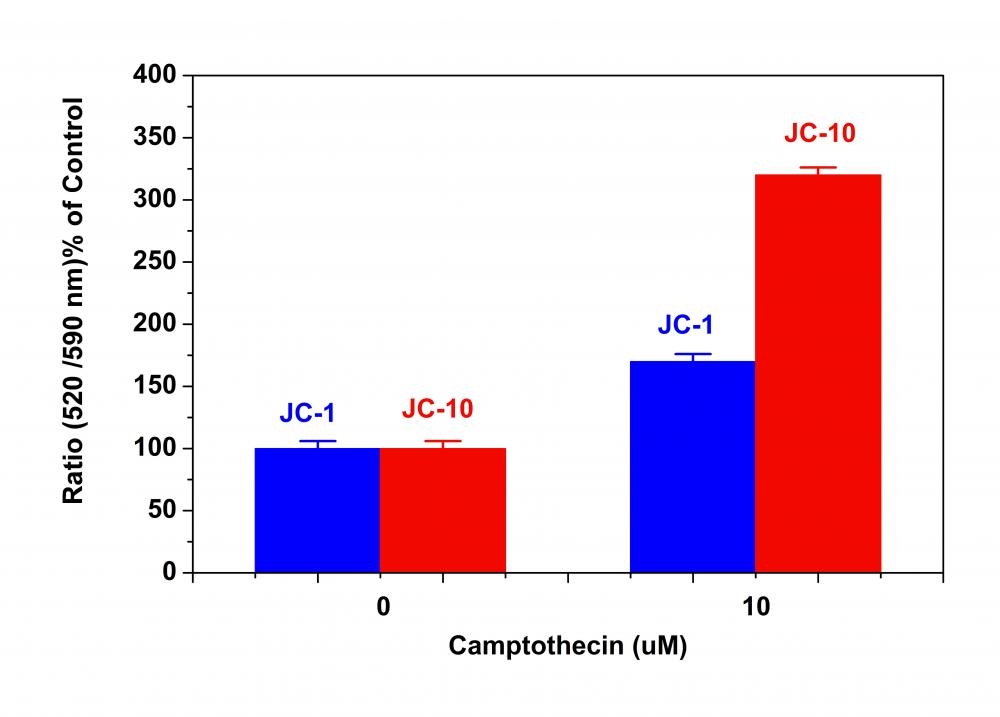
Figure 1. Campotothecin-induced mitochondria membrane potential changes were measured with JC-10™ and JC-1 in Jurkat cells. After Jurkat cells were treated with camptothecin (10 µM) for 4 hours, JC-1 and JC-10™ dye working solutions were added to the wells and incubated for 30 minutes. The fluorescence intensities for both J-aggregates and monomeric forms of JC-1 and JC-10™ were measured at Ex/Em = 490/525 nm (Cutoff = 515 nm) and 490/590 nm (Cutoff = 570 nm) with NOVOstar microplate reader (BMG Labtech).
Citations
View all 48 citations: Citation Explorer
Antineoplastic Effects and Mechanisms of a New RGD Chimeric Peptide from Bullfrog Skin on the Proliferation and Apoptosis of B16F10 Cells
Authors: Jiang, Xuan and Zhang, Xin and Fu, Chao and Zhao, Ruili and Jin, Tianming and Liu, Mengyue and Pan, Chenhao and Li, Liu An and Ma, Jifei and Yu, Enyuan and others,
Journal: The Protein Journal (2021): 1--12
Authors: Jiang, Xuan and Zhang, Xin and Fu, Chao and Zhao, Ruili and Jin, Tianming and Liu, Mengyue and Pan, Chenhao and Li, Liu An and Ma, Jifei and Yu, Enyuan and others,
Journal: The Protein Journal (2021): 1--12
Human VAMP3 Suppresses or Negatively Regulates Bax Induced Apoptosis in Yeast
Authors: Akintade, Damilare D and Chaudhuri, Bhabatosh
Journal: Biomedicines (2021): 95
Authors: Akintade, Damilare D and Chaudhuri, Bhabatosh
Journal: Biomedicines (2021): 95
Human VAMP3 Suppresses or Negatively Regulates Bax Induced Apoptosis in Yeast. Biomedicines 2021, 9, 95
Authors: Akintade, DD and Chaudhuri, B
Journal: (2021)
Authors: Akintade, DD and Chaudhuri, B
Journal: (2021)
SIRT3 consolidates heterochromatin and counteracts senescence
Authors: Diao, Zhiqing and Ji, Qianzhao and Wu, Zeming and Zhang, Weiqi and Cai, Yusheng and Wang, Zehua and Hu, Jianli and Liu, Zunpeng and Wang, Qiaoran and Bi, Shijia and others,
Journal: Nucleic acids research (2021): 4203--4219
Authors: Diao, Zhiqing and Ji, Qianzhao and Wu, Zeming and Zhang, Weiqi and Cai, Yusheng and Wang, Zehua and Hu, Jianli and Liu, Zunpeng and Wang, Qiaoran and Bi, Shijia and others,
Journal: Nucleic acids research (2021): 4203--4219
Apoptosis, Induced by Human $\alpha$-Synuclein in Yeast, Can Occur Independent of Functional Mitochondria
Authors: Akintade, Damilare D and Chaudhuri, Bhabatosh
Journal: Cells (2020): 2203
Authors: Akintade, Damilare D and Chaudhuri, Bhabatosh
Journal: Cells (2020): 2203
Modeling Secondary Iron Overload Cardiomyopathy with Human Induced Pluripotent Stem Cell-Derived Cardiomyocytes
Authors: Rhee, June-Wha and Yi, Hyoju and Thomas, Dilip and Lam, Chi Keung and Belbachir, Nadjet and Tian, Lei and Qin, Xulei and Malisa, Jessica and Lau, Edward and Paik, David T and others,
Journal: Cell Reports (2020): 107886
Authors: Rhee, June-Wha and Yi, Hyoju and Thomas, Dilip and Lam, Chi Keung and Belbachir, Nadjet and Tian, Lei and Qin, Xulei and Malisa, Jessica and Lau, Edward and Paik, David T and others,
Journal: Cell Reports (2020): 107886
Identification of proteins involved in transcription/translation (eEF 1A1) as an inhibitor of Bax induced apoptosis
Authors: Akintade, Damilare D and Chaudhuri, Bhabatosh
Journal: Molecular biology reports (2020): 6785--6792
Authors: Akintade, Damilare D and Chaudhuri, Bhabatosh
Journal: Molecular biology reports (2020): 6785--6792
Cellular calcification induced by inorganic polyphosphate involves ATP depletion and opening of the mitochondrial permeability transition pore (mPTP)
Authors: Tsutsumi, Kaori and Sasase, Tatsuya
Journal: FEBS Open Bio (2019)
Authors: Tsutsumi, Kaori and Sasase, Tatsuya
Journal: FEBS Open Bio (2019)
Nonantioxidant tetramethoxystilbene abrogates $\alpha$-synuclein-induced yeast cell death but not that triggered by the Bax or $\beta$A4 peptide
Authors: Derf, Asma and Mudududdla, Ramesh and Akintade, Damilare and Williams, Ibidapo S and Abdullaha, Mohd and Chaudhuri, Bhabatosh and Bharate, Sandip B
Journal: ACS omega (2018): 9513--9532
Authors: Derf, Asma and Mudududdla, Ramesh and Akintade, Damilare and Williams, Ibidapo S and Abdullaha, Mohd and Chaudhuri, Bhabatosh and Bharate, Sandip B
Journal: ACS omega (2018): 9513--9532
Role of Mcl-1 in regulation of cell death in human induced pluripotent stem cell-derived cardiomyocytes in vitro
Authors: Guo, Liang and Eldridge, S and y , undefined and Furniss, Michael and Mussio, Jodie and Davis, Myrtle
Journal: Toxicology and Applied Pharmacology (2018)
Authors: Guo, Liang and Eldridge, S and y , undefined and Furniss, Michael and Mussio, Jodie and Davis, Myrtle
Journal: Toxicology and Applied Pharmacology (2018)
Application notes
A Novel Fluorescent Probe for Imaging and Detecting Hydroxyl Radical in Living Cells
Activation of the Mitochondrial Apoptotic Pathway Produces Reactive Oxygen Species and Oxidative Damage in Hepatocytes That Contribute to Liver Tumorigenesis
Calmodulin antagonists induce cell cycle arrest and apoptosis in vitro and inhibit tumor growth in vivo in human multiple myeloma
Cold exposure lowers energy expenditure at the cellular level
Developing high-content assays for hepatoxicity with iPSC-derived cells