Products
Services
Resources
Selection Guides
About
Cell Navigator® Fluorimetric Lipid Droplet Assay Kit
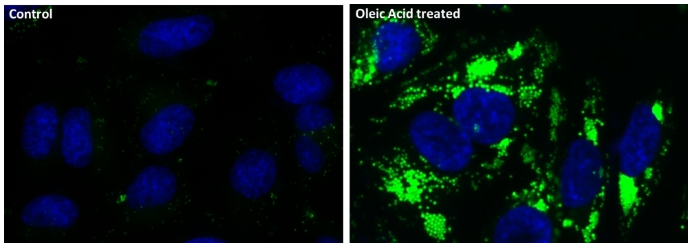
Green Fluorescence
Lipid droplets, also referred to as lipid bodies, oil bodies or adiposomes, are lipid-rich cellular organelles that regulate the storage and hydrolysis of neutral lipids. They also serve as a reservoir of lipid source for many important biological processes such as fatty acid and cellular cholesterol for energy and membrane formation and maintenance. Abnormal accumulation of the cytoplasmic lipid droplets occurs in a variety of pathological conditions and can be an indicator of metabolic deficiency or pathogenesis. AAT Bioquest's Cell Navigator® Fluorimetric Lipid Droplets Assay Kit is a simple assay that could quantitatively measure lipid droplet accumulation. Nile Green™ is used in the kit for lipophilic stain. Nile Green™ is intensely fluorescent in a lipid-rich environment while it has minimal fluorescence in aqueous media. It is an excellent vital stain for the detection of intracellular lipid droplets with fluorescence microscopy, flow cytometry or fluorescence microplate reader. Unlike Nile Red which has broad range of fluorescence spectrum, Nile Green™ stains intracellular lipid droplets with green fluorescence only. It can be used with other fluorescence dyes for multicolor staining. The green fluorescence signal could be observed using the filter set of FITC.
| Catalog | Size | Price | Quantity |
|---|---|---|---|
| 22730 | 200 Tests | Price |

Spectral properties
| Correction factor (260 nm) | 0.015 |
| Correction factor (280 nm) | 0.018 |
| Extinction coefficient (cm -1 M -1) | 81000 |
| Excitation (nm) | 504 |
| Emission (nm) | 510 |
Usage and storage
| Intended use | Research Use Only (RUO) |
Instrument settings
| Fluorescence microscope | |
| Excitation | FITC filter set |
| Emission | FITC filter set |
| Recommended plate | Black wall/clear bottom |
| Fluorescence microplate reader | |
| Excitation | 485 nm |
| Emission | 520 nm |
| Cutoff | 510 nm |
| Recommended plate | Black wall/clear bottom |
| Instrument specification(s) | Bottom read mode |
Contact us
| Telephone | |
| Fax | |
| sales@aatbio.com | |
| International | See distributors |
| Bulk request | Inquire |
| Custom size | Inquire |
| Technical Support | Contact us |
| Request quotation | Request |
| Purchase order | Send to sales@aatbio.com |
| Shipping | Standard overnight for United States, inquire for international |
Page updated on July 16, 2026