logo
AAT Bioquest

Cell Navigator® Fluorimetric Lipid Droplet Assay Kit *Red Fluorescence*

Fluorescence images of intracellular lipid droplets in control (Left) and Oleic Acid treated HeLa cells (Right) using Cell Navigator® Lipid Droplets Fluorescence Assay Kit. HeLa cells were incubated with 300 uM of Oleic Acid for 24 hours to induce intracellular lipid droplets formation. After washing with PBS, the cells were labeled with 1X Droplite™ Red and Hoechst 33342  (Cat#17533).
Ordering information
Price
Catalog Number
Unit Size
Quantity
Add to cart
Additional ordering information
Telephone1-800-990-8053
Fax1-800-609-2943
Emailsales@aatbio.com
InternationalSee distributors
Bulk requestInquire
Custom sizeInquire
ShippingStandard overnight for United States, inquire for international
Request quotation
Spectral properties
Absorbance (nm)560
Extinction coefficient (cm -1 M -1)38000
Excitation (nm)559
Emission (nm)635
Quantum yield0.70001
Storage, safety and handling
Certificate of OriginDownload PDF
H-phraseH303, H313, H333
Hazard symbolXN
Intended useResearch Use Only (RUO)
R-phraseR20, R21, R22
UNSPSC12352200
Related products
Cell Navigator® Live Cell RNA Imaging Kit *Green Fluorescence*
Cell Navigator® Live Cell Endoplasmic Reticulum (ER) Staining Kit *Green Fluorescence*
Cell Navigator® Live Cell Endoplasmic Reticulum (ER) Staining Kit *Red Fluorescence*
Cell Navigator® Lysosome Staining Kit *Green Fluorescence with 405 nm Excitation*
Cell Navigator® Lysosome Staining Kit *NIR Fluorescence*
Cell Navigator® Lysosome Staining Kit *Blue Fluorescence*
Cell Navigator® Lysosome Staining Kit *Green Fluorescence*
Cell Navigator® Lysosome Staining Kit *Orange Fluorescence*
Cell Navigator® Lysosome Staining Kit *Red Fluorescence*
Cell Navigator® Lysosome Staining Kit *Deep Red Fluorescence*
Cell Navigator® F-Actin Labeling Kit *Blue Fluorescence*
Cell Navigator® F-Actin Labeling Kit *Green Fluorescence*
Cell Navigator® F-Actin Labeling Kit *Orange Fluorescence*
Cell Navigator® F-Actin Labeling Kit *Red Fluorescence*
Cell Navigator® Mitochondrion Staining Kit *Blue Fluorescence*
Cell Navigator® Mitochondrion Staining Kit *Green Fluorescence*
Cell Navigator® Mitochondrion Staining Kit *Orange Fluorescence*
Cell Navigator® Mitochondrion Staining Kit *Red Fluorescence*
Cell Navigator® Mitochondrion Staining Kit *Deep Red Fluorescence*
Cell Navigator® Mitochondrion Staining Kit *Orange Fluorescence with 405 nm Excitation*
Cell Navigator® Cell Plasma Membrane Staining Kit *Orange Fluorescence*
Cell Navigator® Cell Plasma Membrane Staining Kit *Red Fluorescence*
Cell Navigator® Mitochondrion Staining Kit *NIR Fluorescence*
Cell Navigator® NBD Ceramide Golgi Staining Kit *Green Fluorescence*
Cell Navigator® Live Cell Tubulin Staining Kit
Cell Navigator® Cell Plasma Membrane Staining Kit *Green Fluorescence*
Cell Navigator® Live Cell Endoplasmic Reticulum (ER) Staining Kit *Blue Fluorescence*
Cell Navigator® TMR Ceramide Golgi Staining Kit *Red Fluorescence*
Cell Navigator® CDy6 Mitosis Imaging Kit
Cell Navigator™ Flow Cytometric Exosome Staining Kit
Show More (20)

OverviewpdfSDSpdfProtocol


See also: Mitochondria
Absorbance (nm)
560
Extinction coefficient (cm -1 M -1)
38000
Excitation (nm)
559
Emission (nm)
635
Quantum yield
0.70001
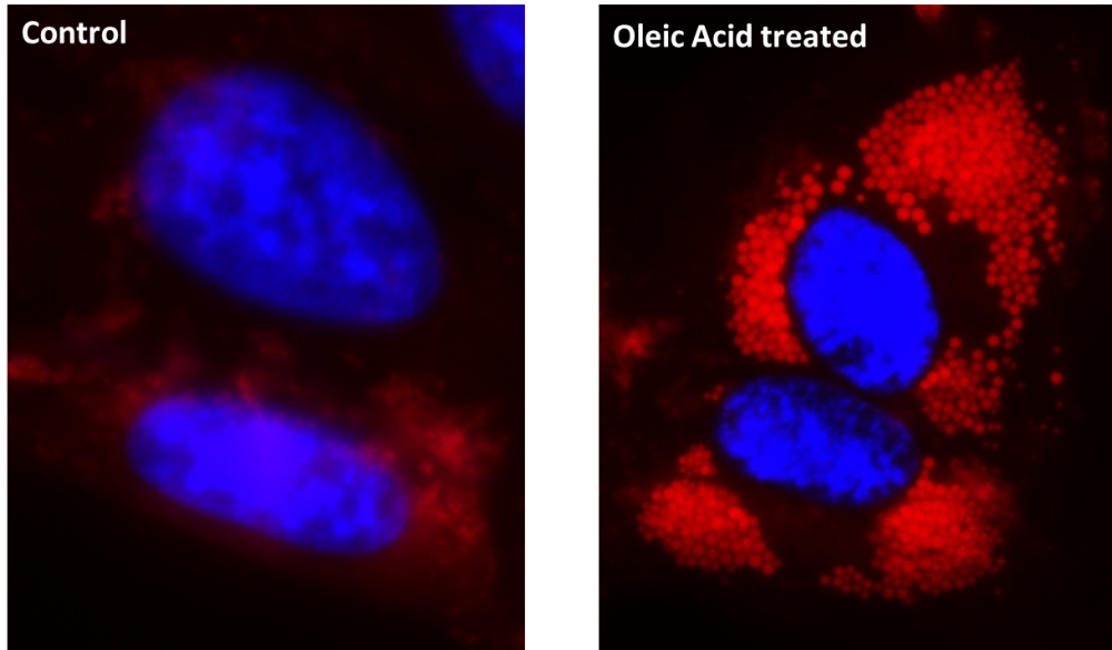
Lipid droplets, also referred to as lipid bodies, oil bodies or adiposomes, are lipid-rich cellular organelles that regulate the storage and hydrolysis of neutral lipids. They also serve as a reservoir of lipid source for many important biological processes such as fatty acid and cellular cholesterol for energy and membrane formation and maintenance. Abnormal accumulation of the cytoplasmic lipid droplets occurs in a variety of pathological conditions and can be an indicator of metabolic deficiency or pathogenesis. AAT Bioquest's Cell Navigator® Fluorimetric Lipid Droplet Assay Kit is a robust tool that could quantitatively measure lipid droplet accumulation. Droplite™ Red is used in the kit for lipophilic stain. Droplite™ Red is intensely fluorescent in a lipid-rich environment while it has minimal fluorescence in aqueous media. It is an excellent vital stain for the detection of intracellular lipid droplets with fluorescence microscopy, flow cytometry or fluorescence microplate reader. The red fluorescence signal could be read observed using the filter set of TRITC.

Platform


Fluorescence microscope

ExcitationTRITC filter set
EmissionTRITC filter set
Recommended plateBlack wall/clear bottom

Fluorescence microplate reader

Excitation550 nm
Emission640 nm
Cutoff610 nm
Recommended plateSolid black

Components


Example protocol


AT A GLANCE

Protocol Summary
  1. Prepare cells with test compounds
  2. Add 100 µL Droplite™ Red staining solution
  3. Incubate at room temperature or 37°C for 10 to 30 min
  4. Read fluorescence intensity at Ex/Em = 550/640 nm (Cutoff = 610 nm), image cells using fluorescence microscope with TRITC filter or flow cytometer with FL1 channel 
Important      Following is our recommended protocol for live cells. This protocol only provides a guideline, and should be modified according to your specific needs. Thaw all the kit components at room temperature before starting the experiment.

CELL PREPARATION

For guidelines on cell sample preparation, please visit https://www.aatbio.com/resources/guides/cell-sample-preparation.html

PREPARATION OF STOCK SOLUTIONS

Unless otherwise noted, all unused stock solutions should be divided into single-use aliquots and stored at -20 °C after preparation. Avoid repeated freeze-thaw cycles.

Droplite™ Red staining solution
Dilute 2 µL of Droplite™ Red (Component A) to 1 mL of Staining Buffer (Component B). Protect from light. Note: 20 µL of Droplite™ Red (Component A) is enough for one 96-well plate. The optimal concentration of the Droplite™ Red varies depending on specific applications. The staining conditions may be modified according to a particular cell type and the permeability of the cells or tissues to the probe.

PREPARATION OF WORKING SOLUTION

Add 2 µL of Droplite™ Red (Component A) to 1 mL of Staining Buffer (Component B) to make Droplite™ Red staining soluton. Protect from light.
Note     20 µL of Droplite™ Red (Component A) is enough for one 96-well plate. The optimal concentration of the Droplite™ Red varies depending on specific applications. The staining conditions may be modified according to a particular cell type and the permeability of the cells or tissues to the probe.

SAMPLE EXPERIMENTAL PROTOCOL

For adherent cells:
  1. Grow cells either in a 96-well black wall/clear bottom plate (100 µL/well/96-well) or on cover-slips inside a petri dish filled with the appropriate culture medium.
  2. Gently aspirate the culture medium, and add equal volume (such as 100 µL/well/96-well plate) of the Droplite™ Red staining solution.
  3. Incubate the cells in a 37 °C, 5% CO2 incubator for 10 - 30 minutes.
  4. Remove Droplite™ Red staining solution (Optional).
  5. Read the fluorescence intensity with a microplate reader at Ex/Em =550/640 nm (Cutoff = 610 nm) or observe the cells using a fluorescence microscope with a TRITC filter set. 

For suspension cells:
  1. Centrifuge the cells at 1000 rpm for 5 minutes to get 1 - 5 × 10⁵ cells per tube.
  2. Resuspend cells in 500 µL of Droplite™ Red staining solution.
  3. Incubate at room temperature or 37 °C for 10 to 30 min, protected from light.
  4. Centrifuge to remove the Droplite™ Red staining solution, and resuspend cells in 500 µL of pre-warmed medium or buffer of your choice to get 1 - 5 × 10⁵ cells per tube (Optional).
  5. Monitor the fluorescence increase using fluorescence microscope with a TRITC filter set or flow cytometer with FL1 channel.
    Note      Since Droplite™ Red has minimal fluorescence in aqueous media, aspiration of the growth medium and removal of Droplite™ Red staining solution after staining is optional. Stained cells can be fixed with 3 - 4% formaldehyde. In addition, prefixed cells (3 - 4% formaldehyde fixation) can be stained with Droplite™ Red staining solution. 

Spectrum


Open in Advanced Spectrum Viewer
spectrum

Spectral properties

Absorbance (nm)560
Extinction coefficient (cm -1 M -1)38000
Excitation (nm)559
Emission (nm)635
Quantum yield0.70001

Product Family


NameExcitation (nm)Emission (nm)Extinction coefficient (cm -1 M -1)Correction Factor (260 nm)Correction Factor (280 nm)
Cell Navigator® Fluorimetric Lipid Droplet Assay Kit *Green Fluorescence*504510810000.0150.018

Images


References


View all 26 references: Citation Explorer
Live cell imaging and analysis of lipid droplets biogenesis in HCV infected cells
Authors: Nevo-Yassaf, I.; Lovelle, M.; Nahmias, Y.; Hirschberg, K.; Sklan, E. H.
Journal: Methods (2017)
Lipid droplets and associated proteins in sebocytes
Authors: Schneider, M. R.
Journal: Exp Cell Res (2016): 205-8
and beyond
Authors: Wang, C. W., Lipid droplets, lipophagy
Journal: Biochim Biophys Acta (2016): 793-805
Acute accumulation of free cholesterol induces the degradation of perilipin 2 and Rab18-dependent fusion of ER and lipid droplets in cultured human hepatocytes
Authors: Makino, A.; Hullin-Matsuda, F.; Murate, M.; Abe, M.; Tomishige, N.; Fukuda, M.; Yamashita, S.; Fujimoto, T.; Vidal, H.; Lagarde, M.; Delton, I.; Kobayashi, T.
Journal: Mol Biol Cell (2016): 3293-3304
Expression of hepatic lipid droplets is decreased in the nitrofen model of congenital diaphragmatic hernia
Authors: Takahashi, H.; Kutasy, B.; Friedmacher, F.; Takahashi, T.; Puri, P.
Journal: Pediatr Surg Int (2016): 155-60
The life cycle of lipid droplets
Authors: Hashemi, H. F.; Goodman, J. M.
Journal: Curr Opin Cell Biol (2015): 119-24
distribution and saturation level in Non-Alcoholic Fatty Liver Disease in mice
Authors: Kochan, K.; Maslak, E.; Krafft, C.; Kostogrys, R.; Chlopicki, S.; Baranska, M., Raman spectroscopy analysis of lipid droplets content
Journal: J Biophotonics (2015): 597-609
Spastin binds to lipid droplets and affects lipid metabolism
Authors: Papadopoulos, C.; Orso, G.; Mancuso, G.; Herholz, M.; Gumeni, S.; Tadepalle, N.; Jungst, C.; Tzschichholz, A.; Schauss, A.; Honing, S.; Trifunovic, A.; Daga, A.; Rugarli, E. I.
Journal: PLoS Genet (2015): e1005149
Structure, function and metabolism of hepatic and adipose tissue lipid droplets: implications in alcoholic liver disease
Authors: Natarajan, S. K.; Rasineni, K.; Ganesan, M.; Feng, D.; McVicker, B. L.; McNiven, M. A.; Osna, N. A.; Mott, J. L.; Casey, C. A.; Kharb and a, K. K.
Journal: Curr Mol Pharmacol (2015)
Chemical imaging of lipid droplets in muscle tissues using hyperspectral coherent Raman microscopy
Authors: Billecke, N.; Rago, G.; Bosma, M.; Eijkel, G.; Gemmink, A.; Leproux, P.; Huss, G.; Schrauwen, P.; Hesselink, M. K.; Bonn, M.; Parekh, S. H.
Journal: Histochem Cell Biol (2014): 263-73