Products
Services
Resources
Selection Guides
About
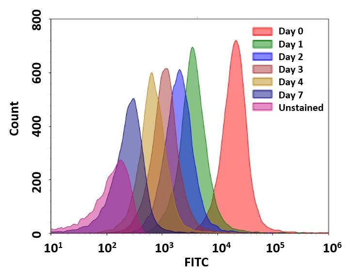
CytoTell® UltraGreen
CytoTell® UltraGreen is a green fluorescent cell proliferation dye for monitoring cell division and long-term tracking of live cells in flow cytometry and fluorescence microscopy applications.
- Reduced cytotoxicity: Provides lower cellular toxicity for long-term live cell proliferation studies
- Enhanced proliferation tracking: Enables monitoring of successive cell generations through progressive fluorescence dilution
- 488 nm laser compatible: Optimized for excitation with blue laser-based flow cytometers and imaging systems
- Improved cellular retention: Designed for enhanced intracellular retention during long-term cell tracking applications
- Broad application compatibility: Suitable for flow cytometry, fluorescence microscopy, and long-term live cell tracking applications
| Catalog | Size | Price | Quantity |
|---|---|---|---|
| 22240 | 500 Tests | Price | |
| 22241 | 2x500 Tests | Price |

Physical properties
| Molecular weight | ~500 |
| Solvent | DMSO |
Spectral properties
| Excitation (nm) | 492 |
| Emission (nm) | 514 |
Usage and storage
| Intended use | Research Use Only (RUO) |
| Storage | Freeze (< -15 °C); Minimize light exposure |
Instrument settings
| Flow cytometer | |
| Excitation | 488 nm laser |
| Emission | 530/30 nm filter |
| Instrument specification(s) | FITC channel |
Contact us
| Telephone | |
| Fax | |
| sales@aatbio.com | |
| International | See distributors |
| Bulk request | Inquire |
| Custom size | Inquire |
| Technical Support | Contact us |
| Request quotation | Request |
| Purchase order | Send to sales@aatbio.com |
| Shipping | Standard overnight for United States, inquire for international |
Page updated on July 30, 2026