Helixyte™ Green dsDNA Quantifying Reagent *200X DMSO Solution*
Price | |
Catalog Number | |
Unit Size | |
Quantity |
Telephone | 1-800-990-8053 |
Fax | 1-800-609-2943 |
sales@aatbio.com | |
Quotation | Request |
International | See distributors |
Shipping | Standard overnight for United States, inquire for international |
Molecular weight | 661.17 |
Solvent | DMSO |
Excitation (nm) | 502 |
Emission (nm) | 522 |
H-phrase | H303, H313, H340 |
Hazard symbol | T |
Intended use | Research Use Only (RUO) |
R-phrase | R20, R21, R68 |
Storage | Freeze (< -15 °C); Minimize light exposure |
UNSPSC | 41116134 |
Overview | ![]() ![]() |
Molecular weight 661.17 | Excitation (nm) 502 | Emission (nm) 522 |
Platform
Fluorescence microplate reader
Excitation | 490 |
Emission | 525 |
Cutoff | 515 |
Recommended plate | Solid black |
Example protocol
PREPARATION OF WORKING SOLUTION
The following example is a protocol for quantifying dsDNA with Helixyte Green™. Before opening, allow the vial of Helixyte Green™ to warm to room temperature.
Note: Exercise caution when working with Helixyte Green™ dsDNA stain, as there is currently no available data regarding its potential mutagenicity or toxicity. Due to its affinity for nucleic acids, this reagent should be handled as a potential mutagen and with appropriate care. Additionally, take special care when handling the DMSO stock solution, as DMSO has been shown to facilitate the entry of organic molecules into tissues.
- To prepare an aqueous working solution of Helixyte Green™, dilute the concentrated DMSO solution 200-fold in TE buffer (10 mM Tris-HCl, 1 mM EDTA, pH 7.5-8.0). For example, adding 50 μL of Helixyte Green™ to 10 mL of TE will prepare enough working solution to assay 100 samples in a final volume of 200 µL. Protect the working solution from light by covering it with foil or placing it in the dark.Note: We recommend preparing this solution in a plastic container rather than glass, as the dye may adsorb to glass surfaces.Note: For best results, this solution should be used within a few hours of its preparation.
SAMPLE EXPERIMENTAL PROTOCOL
Prepare a 1 mg/mL stock solution of dsDNA (such as calf thymus DNA from Sigma) in ddH2O
To prepare a 10 μg/mL dsDNA solution, add 10 μL of the 1 mg/mL dsDNA stock solution (from Step 2.1) to 990 µL of TE buffer. Next, perform 1:10 and 1:2 serial dilutions to obtain concentrations of 1000, 100, 50, 25, 12.5, 6.25, 3.125, and 0 ng/mL.
Add the dsDNA standards and test samples containing DNA into a solid black 96-well microplate, following the instructions provided in Tables 1 and 2.
Table 1. The layout of dsDNA standards and test samples in a solid black 96-well microplate.*
BL | BL | TS | TS | .... | .... |
|
|
|
|
|
|
DS1 | DS1 | .... | .... | .... | .... |
|
|
|
|
|
|
DS2 | DS2 | .... | .... | .... | .... |
|
|
|
|
|
|
DS3 | DS3 |
|
|
|
|
|
| ||||
DS4 | DS4 |
|
|
|
|
|
| ||||
DS5 | DS5 |
|
|
|
|
|
| ||||
DS6 | DS6 |
|
|
|
|
|
| ||||
DS7 | DS7 |
|
|
|
|
|
|
*Note: DS = dsDNA Standards; BL= Blank Control; TS =Test Samples
Table 2. Reagent composition for each well.*
dsDNA Standard | Blank Control | Test Sample |
Serial dilutions* (100 µL) | TE: 100 μL | 100 μL |
*Note: Add the serial dilutions of dsDNA standards from 0.1 to 1000 ng/mL into wells from DS1 to DS7 in duplicate.
Add 100 μL of dsDNA assay mixture (from Step 1.1) to each well of the dsDNA standard, blank control, and test samples (see Step 2.3) to make the total dsDNA assay volume of 200 µL/well.
Note: For a 384-well plate, add 25 μL sample and 25 μL of dsDNA assay mixture per well.
Note: For cuvette-based assays, add 1mL sample and 1mL of dsDNA assay mixture per cuvette.
Incubate the reaction at room temperature for 5 to 10 minutes, protected from light.
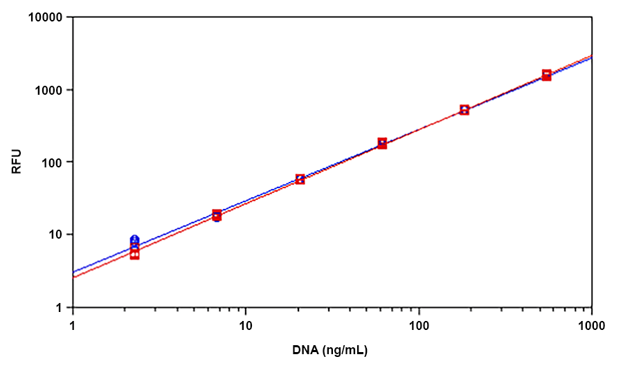
Monitor the fluorescence increase with a fluorescence microplate reader at Ex/Em = 490/525 nm, cut off at 515 nm.
The fluorescence obtained in the blank wells (containing only TE buffer) is used as a control and will be subtracted from the values obtained in the wells containing dsDNA reactions. The DNA concentrations of the test samples can be determined by referencing the standard curve generated in the DNA Standard Curve step.
Calculators
Common stock solution preparation
0.1 mg | 0.5 mg | 1 mg | 5 mg | 10 mg | |
1 mM | 151.247 µL | 756.235 µL | 1.512 mL | 7.562 mL | 15.125 mL |
5 mM | 30.249 µL | 151.247 µL | 302.494 µL | 1.512 mL | 3.025 mL |
10 mM | 15.125 µL | 75.624 µL | 151.247 µL | 756.235 µL | 1.512 mL |
Molarity calculator
Mass (Calculate) | Molecular weight | Volume (Calculate) | Concentration (Calculate) | Moles | ||||
/ | = | x | = |
Images
Citations
Authors: Lee, Kathleen and Jackson, Anisha and John, Nikita and Zhang, Ryan and Ozhava, Derya and Bhatia, Mohit and Mao, Yong
Journal: Journal of Functional Biomaterials (2023): 218
Authors: Salleh, Mohd Hairul Mohd and Esa, Yuzine and Ngalimat, Mohamad Syazwan and Chen, Pelf Nyok
Journal: PeerJ (2022): e12970
Authors: Fatma, Yuli and RUSMANA, IMAN and WAHYUDI, ARIS TRI and others,
Journal: Biodiversitas Journal of Biological Diversity (2019)
References
Authors: Peterson EJ, Kireev D, Moon AF, Midon M, Janzen WP, Pingoud A, Pedersen LC, Singleton SF.
Journal: J Biomol Screen (2013): 247
Authors: Chen Y, Sonnaert M, Roberts SJ, Luyten FP, Schrooten J.
Journal: Tissue Eng Part C Methods (2012): 444
Authors: Dragan AI, Casas-Finet JR, Bishop ES, Strouse RJ, Schenerman MA, Geddes CD.
Journal: Biophys J (2010): 3010
Authors: Abiodun OO, Gbotosho GO, Ajaiyeoba EO, Happi CT, Hofer S, Wittlin S, Sowunmi A, Brun R, Oduola AM.
Journal: Parasitol Res (2010): 933
Authors: Dragan AI, Bishop ES, Casas-Finet JR, Strouse RJ, Schenerman MA, Geddes CD.
Journal: J Immunol Methods (2010): 95
Authors: Moreno LA, Cox KL.
Journal: J Vis Exp. (2010)
Authors: Holden MJ, Haynes RJ, Rabb SA, Satija N, Yang K, Blasic JR, Jr.
Journal: J Agric Food Chem (2009): 7221
Authors: Ikeda Y, Iwakiri S, Yoshimori T.
Journal: J Pharm Biomed Anal (2009): 997
Authors: Noothi SK, Kombrabail M, Kundu TK, Krishnamoorthy G, Rao BJ.
Journal: FEBS J (2009): 541
Authors: Ren X, Xu QH.
Journal: Langmuir (2009): 43