Protonex™ Green 500 Dextran
Ordering information
Price | |
Catalog Number | |
Unit Size | |
Quantity |
Additional ordering information
Telephone | 1-800-990-8053 |
Fax | 1-800-609-2943 |
sales@aatbio.com | |
Quotation | Request |
International | See distributors |
Shipping | Standard overnight for United States, inquire for international |
Physical properties
Molecular weight | ~11000 |
Solvent | Water |
Spectral properties
Extinction coefficient (cm -1 M -1) | 4000 |
Excitation (nm) | 445 |
Emission (nm) | 503 |
Storage, safety and handling
H-phrase | H303, H313, H333 |
Hazard symbol | XN |
Intended use | Research Use Only (RUO) |
R-phrase | R20, R21, R22 |
Storage | Freeze (< -15 °C); Minimize light exposure |
UNSPSC | 12352200 |
Overview | ![]() ![]() |
See also: Intracellular pH, Physiological Probes
Molecular weight ~11000 | Extinction coefficient (cm -1 M -1) 4000 | Excitation (nm) 445 | Emission (nm) 503 |
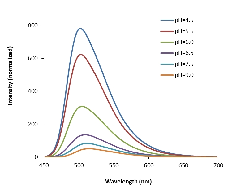
Protonex™ Green dye demonstrated pH-dependent fluorescence. Unlike most of the existing fluorescent dyes that are more fluorescent at higher pH, acidic conditions enhance the fluorescence of Protonex™ Green dye. The fluorescence of Protonex™ Green dye increases as pH decreases from neutral to the acidic. The lack of fluorescence outside the cell eliminates the wash steps. Protonex™ Green dye provides a powerful tool to monitor acidic cell compartments such as endosomes and lysosomes. Protonex™ Green dye is non-fluorescent outside the cells, but fluoresces brightly green in acidic compartments (such as phagosomes, lysosomes and endosomes). This Protonex™ Green enables the specific detection of cellular acidic compartments with reduced signal variability and improved accuracy for imaging or flow applications. Protonex™ Green has the spectral properties similar to those of FITC, making the common filter set of FITC readily available to the assays of Protonex™ Green.
Spectrum
Open in Advanced Spectrum Viewer


Spectral properties
Extinction coefficient (cm -1 M -1) | 4000 |
Excitation (nm) | 445 |
Emission (nm) | 503 |
Product Family
Name | Excitation (nm) | Emission (nm) | Extinction coefficient (cm -1 M -1) |
Protonex™ Green 500, SE | 445 | 503 | 4000 |
Citations
View all 1 citations: Citation Explorer
PHD2 is a regulator for glycolytic reprogramming in macrophages
Authors: Guentsch, Annemarie and Beneke, Angelika and Swain, Lija and Farhat, Katja and Nagarajan, Shunmugam and Wielockx, Ben and Raithatha, Kaamini and Dudek, Jan and Rehling, Peter and Zieseniss, Anke and others, undefined
Journal: Molecular and Cellular Biology (2016): MCB--00236
Authors: Guentsch, Annemarie and Beneke, Angelika and Swain, Lija and Farhat, Katja and Nagarajan, Shunmugam and Wielockx, Ben and Raithatha, Kaamini and Dudek, Jan and Rehling, Peter and Zieseniss, Anke and others, undefined
Journal: Molecular and Cellular Biology (2016): MCB--00236
References
View all 56 references: Citation Explorer
Monitoring phospholipid dynamics during phagocytosis: application of genetically-encoded fluorescent probes
Authors: Sarantis H, Grinstein S.
Journal: Methods Cell Biol (2012): 429
Authors: Sarantis H, Grinstein S.
Journal: Methods Cell Biol (2012): 429
Phagocytosis and digestion of pH-sensitive fluorescent dye (Eos-FP) transfected E. coli in whole blood assays from patients with severe sepsis and septic shock
Authors: Schreiner L, Huber-Lang M, Weiss ME, Hohmann H, Schmolz M, Schneider EM.
Journal: J Cell Commun Signal (2011): 135
Authors: Schreiner L, Huber-Lang M, Weiss ME, Hohmann H, Schmolz M, Schneider EM.
Journal: J Cell Commun Signal (2011): 135
The application of fluorescent probes for the analysis of lipid dynamics during phagocytosis
Authors: Flannagan RS, Grinstein S.
Journal: Methods Mol Biol (2010): 121
Authors: Flannagan RS, Grinstein S.
Journal: Methods Mol Biol (2010): 121
Quantification of microsized fluorescent particles phagocytosis to a better knowledge of toxicity mechanisms
Authors: Leclerc L, Boudard D, Pourchez J, Forest V, Sabido O, Bin V, Palle S, Grosseau P, Bernache D, Cottier M.
Journal: Inhal Toxicol (2010): 1091
Authors: Leclerc L, Boudard D, Pourchez J, Forest V, Sabido O, Bin V, Palle S, Grosseau P, Bernache D, Cottier M.
Journal: Inhal Toxicol (2010): 1091
Analysis of macrophage phagocytosis: quantitative assays of phagosome formation and maturation using high-throughput fluorescence microscopy
Authors: Steinberg BE, Grinstein S.
Journal: Methods Mol Biol (2009): 45
Authors: Steinberg BE, Grinstein S.
Journal: Methods Mol Biol (2009): 45
Phagocytosis and postphagocytic reaction of cord blood and adult blood monocyte after infection with green fluorescent protein-labeled Escherichia coli and group B Streptococci
Authors: Gille C, Leiber A, Mundle I, Spring B, Abele H, Spellerberg B, Hartmann H, Poets Ch F, Orlikowsky TW.
Journal: Cytometry B Clin Cytom (2009): 271
Authors: Gille C, Leiber A, Mundle I, Spring B, Abele H, Spellerberg B, Hartmann H, Poets Ch F, Orlikowsky TW.
Journal: Cytometry B Clin Cytom (2009): 271
A fluorescently tagged C-terminal fragment of p47phox detects NADPH oxidase dynamics during phagocytosis
Authors: Li XJ, Tian W, Stull ND, Grinstein S, Atkinson S, Dinauer MC.
Journal: Mol Biol Cell (2009): 1520
Authors: Li XJ, Tian W, Stull ND, Grinstein S, Atkinson S, Dinauer MC.
Journal: Mol Biol Cell (2009): 1520
Analysis of phosphoinositide dynamics during phagocytosis using genetically encoded fluorescent biosensors
Authors: Cosio G, Grinstein S.
Journal: Methods Mol Biol (2008): 287
Authors: Cosio G, Grinstein S.
Journal: Methods Mol Biol (2008): 287
Development of a highly specific rhodamine-based fluorescence probe for hypochlorous acid and its application to real-time imaging of phagocytosis
Authors: Kenmoku S, Urano Y, Kojima H, Nagano T.
Journal: J Am Chem Soc (2007): 7313
Authors: Kenmoku S, Urano Y, Kojima H, Nagano T.
Journal: J Am Chem Soc (2007): 7313
The nonopsonic allogeneic cell phagocytosis of macrophages detected by flow cytometry and two photon fluorescence microscope
Authors: Liu GW, Ma HX, Wu Y, Zhao Y.
Journal: Transpl Immunol (2006): 220
Authors: Liu GW, Ma HX, Wu Y, Zhao Y.
Journal: Transpl Immunol (2006): 220
Application notes
Cell Loading Protocol For Fluorescent pH Indicator, BCECF-AM
A Comparison of Fluorescent Red Calcium Indicators for Detecting Intracellular Calcium Mobilization in CHO Cells
A Meta-Analysis of Common Calcium Indicators
A New Red Fluorescent & Robust Screen Quest™ Rhod-4™ Ca2+Indicator for Screening GPCR & Ca2+ Channel Targets
A New Robust No-Wash FLIPR Calcium Assay Kit for Screening GPCR and Calcium Channel Targets
FAQ
How to measure intracellular pH using flow cytometry?
I am performing a phagocytosis assay of macrophages engulfing Protonex Red 600 labeled tumor cells. How do you recommend I label my cells?
What is an AM ester?
Are NADH and ROS related?
Are there any alternatives for ethidium bromide in agarose gels?
I am performing a phagocytosis assay of macrophages engulfing Protonex Red 600 labeled tumor cells. How do you recommend I label my cells?
What is an AM ester?
Are NADH and ROS related?
Are there any alternatives for ethidium bromide in agarose gels?