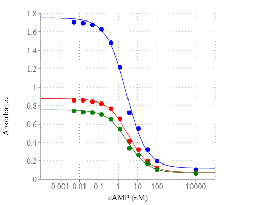
Screen Quest™ Colorimetric ELISA cAMP Assay Kit
Price | |
Catalog Number | |
Unit Size | |
Quantity |
Telephone | 1-800-990-8053 |
Fax | 1-800-609-2943 |
sales@aatbio.com | |
International | See distributors |
Bulk request | Inquire |
Custom size | Inquire |
Shipping | Standard overnight for United States, inquire for international |
H-phrase | H303, H313, H333 |
Hazard symbol | XN |
Intended use | Research Use Only (RUO) |
R-phrase | R20, R21, R22 |
UNSPSC | 12352200 |
Overview | ![]() ![]() |
Platform
Absorbance microplate reader
Absorbance | 405, 650, or 740 nm |
Recommended plate | Clear plate (Component F) |
Components
Example protocol
AT A GLANCE
Protocol summary
- Prepare samples
- Add 75 µL/well of cAMP standard or test samples into the anti-cAMP coated 96-well plate
- Incubate at room temperature for 5-10 mins
- Add 25 µL/well of 1X HRP-cAMP Conjugate
- Incubate at room temperature for 3 hours
- Wash 4 times with 200 µL/well Washing Buffer
- Add 100 µL/well of Amplite™ Green
- Incubate at room temperature for 1 to 3 hours
- Monitor absorbance increase at 405, 650 or 740 nm
Important notes
Do not freeze Anti-cAMP Ab Pre-coated 96-well plate (Component F), store it at 4°C. Allow all the kit components to warm to room temperature before using them. Some material might be stick to the vial cap during the shipment. Briefly centrifuge the vial to collect all the content.
PREPARATION OF STOCK SOLUTION
1. cAMP stock solution (100 µM):
Add 1 mL of Assay Buffer (Component B) to the vial of cAMP Standard (Component A).
2. HRP-cAMP conjugate stock solution (50X):
Add 55 µL (Cat. # 36370) or 550 µL (Cat. # 36371) of Assay Buffer (Component B) into the vial of HRP-cAMP Conjugate (Component C). Note: The unused 50X HRP-cAMP conjugate stock solution should be divided into single use aliquots and stored them at -20 oC.
3. Washing solution (1X):
Add 1 mL of 10X Wash Solution (Component D) to 9 mL distilled water.
PREPARATION OF STANDARD SOLUTION
For convenience, use the Serial Dilution Planner: https://www.aatbio.com/tools/serial-dilution/36370
Make 1:10, 1:100 and 1:3 serial dilutions of cAMP standards in Assay Buffer (Component B) to have 10,000, 100, 30, 10, 3, 1, 0.3, 0.1, 0.03, 0.01, and 0.003 nM cAMP diluted solutions. Store on ice or 4°C.
PREPARATION OF WORKING SOLUTION
HRP-cAMP Conjugate working solution:
Make 1:50 dilution with Assay Buffer (Component B) to have 1X HRP-cAMP conjugate working solution before use. Store it on ice or 4°C. Note: 25 µL of 1X HRP-cAMP conjugate working solution is enough for one assay point; prepare appropriately volume for single use only.
.
For guidelines on cell sample preparation, please visit
https://www.aatbio.com/resources/guides/cell-sample-preparation.html
SAMPLE EXPERIMENTAL PROTOCOL
Prepare samples
- Treat cells as desired:
The following is an example of Hela cells treated with Forskolin to induce cAMP in a 96-well plate format: Aspirate off cell growth medium, add 100 µL/well 100 µM Forskolin in Hanks and 20 mM Hepes buffer (HHBS), incubate in a 5% CO2, 37°C incubator for 15 minutes. Aspirate off cell solution after the incubation, add 100 µL/well of Cell Lysis Buffer (Component E), and incubate at room temperature for another 10 minutes. This cell lysate can be assayed directly or after diluted in Assay Buffer (Component B). Note: Each cell line should be evaluated on an individual basis to determine the optimal cell density. Cells may be seeded the day before or on the day of the experiment depending upon the cell type and/or the effect of the test compounds. - Tissue Samples:
It is important to rapidly freeze tissues after collection (e.g., using liquid nitrogen) due to quick metabolism of cyclic nucleotides in tissue. Weigh the frozen tissue and add 10 - 20 µL/mg of cell lysis buffer. Homogenize the sample on ice. Spin at top speed for 5 minutes and collect the supernatant. The supernatant may be assayed directly. - Urine, Plasma and Culture Medium Samples:
Urine and plasma may be tested directly with 1:200 to 1:1000 dilutions in 1X Lysis Buffer. Culture medium can also be tested with 1:10 to 1:200 dilutions in Lysis Buffer. Note: RPMI medium may contain > 350 fmol/µL cAMP.
cAMP assay
- All the assay wells will be prepared in the following orders: A) cAMP standards, control, or tests samples; B) HRP-cAMP Conjugate.
- Add 75 µL/well of the cAMP diluted standard solution and test samples into each well of the anti-cAMP Ab coated 96-well plate (Component F). We recommended duplicating the assays for each standard and testing sample. Incubate at room temperature for 5 to 10 minutes.
- Add 25 µL/well of 1X HRP-cAMP Conjugate working solution. Incubate at room temperature for 3 hours by placing the plate on shaker.
- Aspirate plate contents, and wash 4 times with 200 µL/well of 1X wash solution.
- Add 100 µL/well of Amplite™ Green (Component G) into each well, and incubate at room temperature for 60 mins to 3 hours, protected from light.
- Monitor the absorbance increase at 405 nm, 650 nm, or 740 nm using an absorbance plate reader.
Images


![Reduction of BTEC migration is dependent on [cAMP]<sub>i</sub> increase. (A) cAMP production upon stimulation with different purinergic agonists or CPA. ATP 100 μM and other antimigratory purinergic agonists induce a strong increase in intracellular cAMP level after 15′ of treatment, as shown in table A (data from at least three independent experiments are normalized to the control). (B) High cAMP levels are able to reduce BTEC migration in wound healing assay. Different inducers of intracellular cAMP increase were used: Forskolin 10 μM (FK), 8-Br-cAMP 500 μM and IBMX 100 μM. Data are normalized on positive CNTRL at 8 hrs and expressed as mean ± S.E.M. Wilcoxon test: *p < 0.001 vs. CNTRL; §p < 0.001 vs. ATP 100 μM; #p < 0.001. (C,i) Actin and paxillin staining in cells treated with FK 10 μM or not (CNTRL) for 5′. Arrows in (C,i) indicate actin filaments distribution and phalloidin fluorescence intensity was evaluated along the major cell axis (white lines in the figures). Scale bar = 10 μm (C,ii) Quantification of the cortical actin localization. (C,iii) Paxillin density quantification upon FK treatment. (C,iv) Total area measured for each condition. Data obtained from three independent experiments are expressed as mean ± S.E.M. Wilcoxon test: *p < 0.001 vs. CNTRL. (D) Effect on BTEC migration (wound healing) of different adenylyl cyclase modulators (ddAdo, NaHCO3), EPAC-1 activator (8-CPT) and PKA inhibitor (H89). Dotted line represents ATP-induced inhibition. Data are normalized on positive CNTRL at 8 hrs and expressed as mean ± S.E.M. Wilcoxon test: *p < 0.001 vs. CNTRL; §p < 0.001 vs. ATP 100 μM; #p < 0.001. Source: <strong>Activation of P2X7 and P2Y11 purinergic receptors inhibits migration and normalizes tumor-derived endothelial cells <em>via</em> cAMP signaling </strong>by Avanzato et al., <em>Scientific Reports</em>, Sept. 2016.](/_next/image?url=https%3A%2F%2Fimages.aatbio.com%2Fproducts%2Ffigures-and-data%2Fscreen-quest-colorimetric-elisa-camp-assay-kit%2Ffigure-for-screen-quest-colorimetric-elisa-camp-assay-kit_johxH.jpg&w=3840&q=75)
Citations
Authors: Avanzato, D and Genova, T and Pla, A Fiorio and Bernardini, M and Bianco, S and Bussolati, B and Mancardi, D and Giraudo, E and Maione, F and Cassoni, P and others, undefined
Journal: Scientific Reports (2016)
Authors: Tomankova, Hana and Valuskova, Paulina and Varejkova, Eva and Rotkova, Jana and Benes, Jan and Myslivecek, Jaromir
Journal: Stress (2015)
Authors: Valu{\v{s}}kov{\'a}, Paulina and Tomankov{\'a}, Hana and Rotkov{\'a}, Jana and Va{\v{r}}ejkov{\'a}, Eva and Bene{\v{s}}, Jan
References
Authors: Rodriguez P, Rojas J.
Journal: J Cell Biochem (2016): 741
Authors: Alqurashi M, Gehring C, Marondedze C.
Journal: Int J Mol Sci (2016): 852
Authors: Ravnskjaer K, Madiraju A, Montminy M.
Journal: Handb Exp Pharmacol (2016): 29
Authors: Miazzi F, Hansson BS, Wicher D.
Journal: J Exp Biol (2016): 1798
Authors: Wang Z, Liu D, Varin A, Nicolas V, Courilleau D, Mateo P, Caubere C, Rouet P, Gomez AM, V and ecasteele G, Fischmeister R, Brenner C.
Journal: Cell Death Dis (2016): e2198
Authors: Almahariq M, Mei FC, Cheng X.
Journal: Acta Biochim Biophys Sin (Shanghai) (2016): 75
Authors: Vedel L, Brauner-Osborne H, Mathiesen JM.
Journal: J Biomol Screen (2015): 849
Authors: Froese A, Nikolaev VO.
Journal: Front Pharmacol (2015): 172
Authors: Zoccarato A, Surdo NC, Aronsen JM, Fields LA, Mancuso L, Dodoni G, Stangherlin A, Livie C, Jiang H, Sin YY, Gesellchen F, Terrin A, Baillie GS, Nicklin SA, Graham D, Szabo-Fresnais N, Krall J, V and eput F, Movsesian M, Furlan L, Corsetti V, Hamilton G, Lefkimmiatis K, Sjaastad I, Zaccolo M.
Journal: Circ Res (2015): 707
Authors: Saegusa Y, Yoshimura K.
Journal: Cytoskeleton (Hoboken) (2015): 412