Gelite™ Safe DNA Gel Stain *GelRed Alternative, 10,000X in water*

| Price | |
| Catalog Number | |
| Unit Size | |
| Quantity |
| Telephone | 1-800-990-8053 |
| Fax | 1-800-609-2943 |
| sales@aatbio.com | |
| International | See distributors |
| Bulk request | Inquire |
| Custom size | Inquire |
| Shipping | Standard overnight for United States, inquire for international |
| Molecular weight | N/A |
| Solvent | Water |
| Absorbance (nm) | 509 |
| Excitation (nm) | 513 |
| Emission (nm) | 552 |
| H-phrase | H303, H313, H333 |
| Hazard symbol | XN |
| Intended use | Research Use Only (RUO) |
| R-phrase | R20, R21, R22 |
| Storage | Freeze (< -15 °C); Minimize light exposure |
| Overview |
Molecular weight N/A | Absorbance (nm) 509 | Excitation (nm) 513 | Emission (nm) 552 |
Platform
Gel Imager
| Excitation | UV Transilluminator/Blue laser |
| Emission | SYBR® filter, GelStar® filter, GelGreen® filter, or GelRed® filter |
Example protocol
PREPARATION OF WORKING SOLUTION
Make 1X Gelite™ Safe working solution by diluting the 10,000X stock reagent with a buffer of your choice in a pH range of 7.5-8.5 (e.g., TAE, TBE, or TE, preferably pH 8.2).
Note: Staining solutions prepared in water are less stable than those prepared in buffer and must be used within 24 hours to ensure maximal staining sensitivity.
SAMPLE EXPERIMENTAL PROTOCOL
The following protocols are recommended. However, some comparisons might be made to determine which one better meets your needs.
Run gels according to your standard protocol.
Place the gel in a suitable polypropylene container. Gently add a sufficient amount of the 1X staining solution to submerge the gel.
Note: Do not use a glass container, as it will adsorb much of the dye in the staining solution.
Agitate the gel gently at room temperature for ~30 to 60 minutes. Protect the staining container from light.
Note: Destaining is not required. Image can be acquired without any wash steps.
Image the gel with a 300 nm/254 nm ultraviolet transilluminator or a laser-based gel scanner using a long path green filter such as a SYBR® filter, GelStar® filter, GelGreen® filter, or GelRed® filter.
Prepare agarose gel solution using your standard protocol.
Dilute the 10,000X Gelite™ Safe stock reagent into the gel solution at 1:10,000 just prior to pouring the gel and mix thoroughly.
Run gels according to your standard protocol.
Image the gel with a 300 nm/254 nm ultraviolet transilluminator or a laser-based gel scanner using a long path green filter such as a SYBR® filter, GelStar® filter, GelGreen® filter, or GelRed® filter.
Spectrum
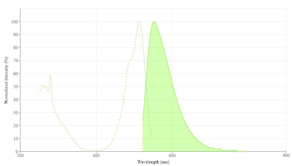
Spectral properties
| Absorbance (nm) | 509 |
| Excitation (nm) | 513 |
| Emission (nm) | 552 |
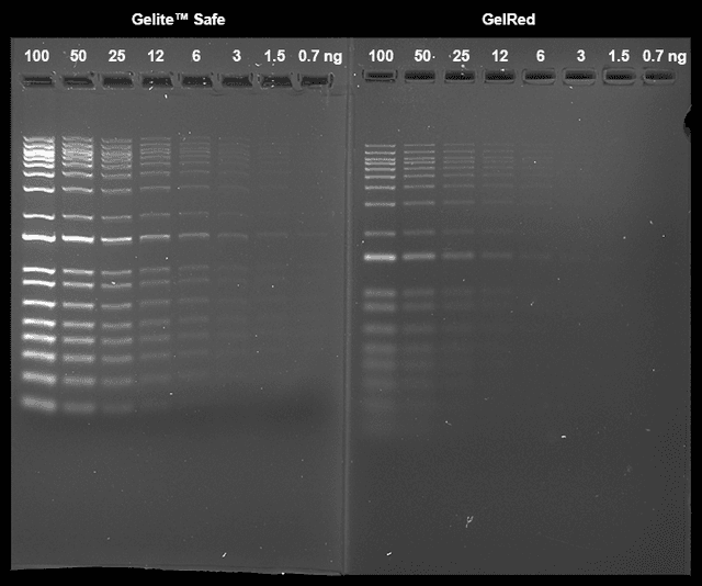
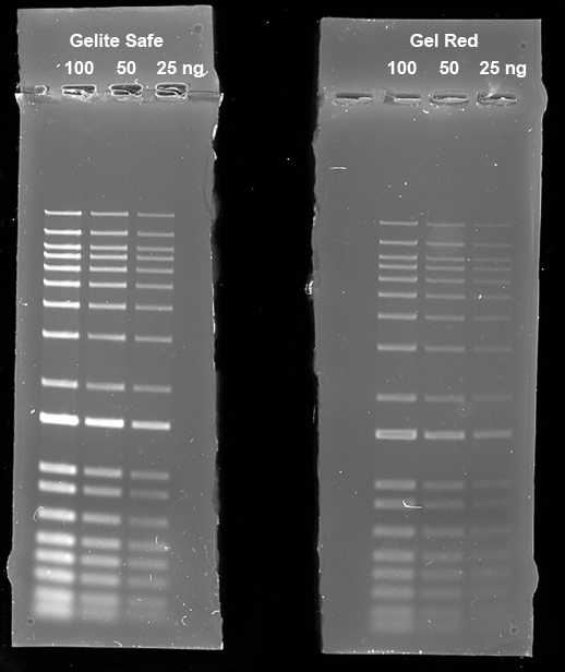
Images

Citations
Authors: Esquivel, Maria and Fernando, Johann and Fisher, Anna and Leong, Cameron and Weaver, Adam
Journal: (2022)
Authors: Nguyen, Kim Cuc Thi and Truong, Phuc Hung and Thi, Hoa Truong and Ho, Xuan Tuy and Van Nguyen, Phu
Application notes
Fluorescent Oligonucleotide Labeling Reagents
Monitoring of Mitochondrial Membrane Potential Changes in Live Cells Using JC-10
Selective Analysis of RNA in Live and Fixed Cells with StrandBrite RNA Green
Cell Loading Protocol For Fluorescent pH Indicator, BCECF-AM
FAQ
What dye works best for staining and tracking lysosomes in live cells for several hours?
How can I lyse my cells without lysing the nuclear membrane?
Do you have any dual-fluorescence nucleic acid stains that interact with both DNA and RNA?
Do you have any fixable mitochondria staining assay kits?