Nucleic Acid Building Blocks

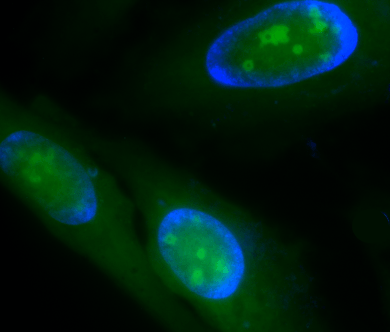
Fluorescence image of RNA staining in live HeLa cells. Cells were stained using Cell Navigator® Live Cell RNA Imaging Kit (Green) and counter-stained with Hoechst 33342 (Blue).
Composition of Nucleic Acids

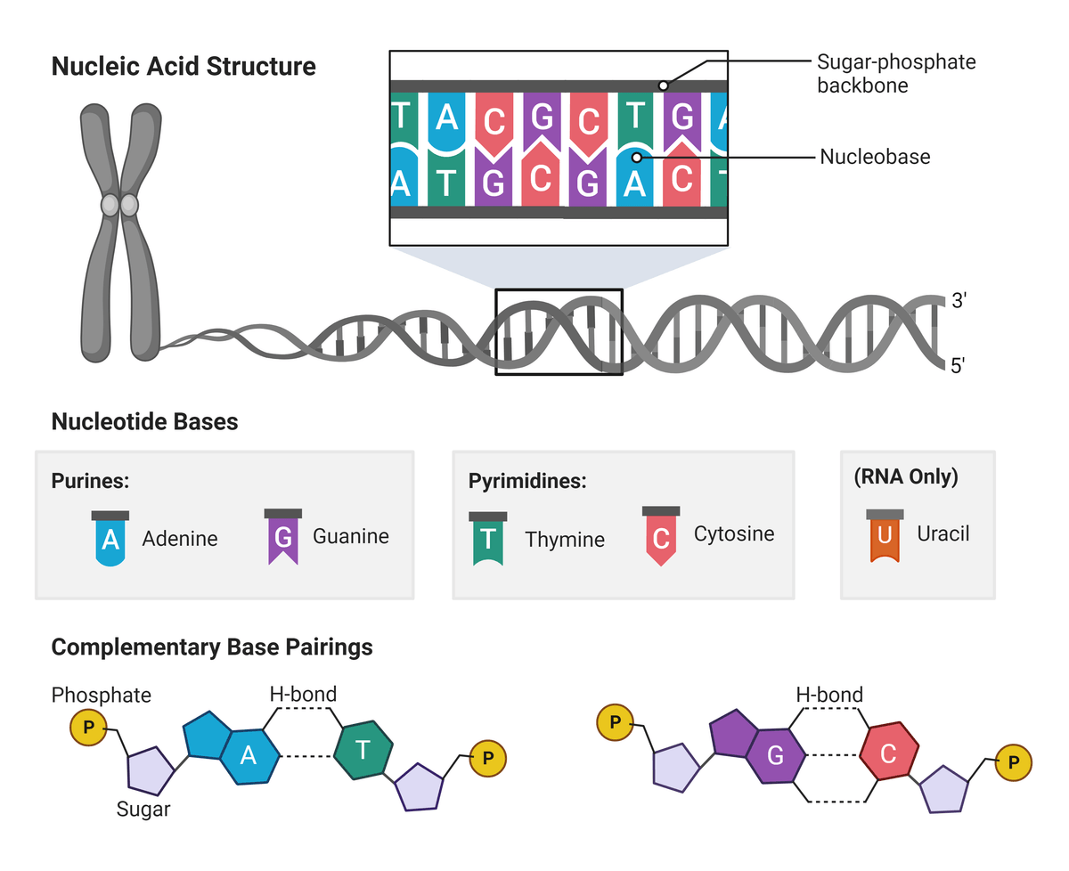
Simplified overview of nucleotide base links and overarching three-dimensional structure. Figure made in BioRender.
The nitrogen-containing base can be adenine (A) or guanine (G), cytosine (C), thymine (T), or uracil (U). A/G are termed purines for their double ringed structure, and C/T/U are termed pyrimidines for their single ring. Notably, only U is found in RNA which often replaces T molecules. These bases are connected to a pentose sugar that contains 5 carbons numbered 1' to 5'. In DNA this sugar is deoxyribose and is located on the second carbon of the pentose ring (2'). In RNA this sugar is ribose which lacks a 2'-hydroxyl (-OH) group. Phosphate groups connect successive sugar residues by bridging the 5'-hydroxyl group on one sugar to the 3'-hydroxyl group of the next.
Each DNA and RNA strand has a sense of direction, where the 3' carbon of the deoxyribose of one nucleotide will always organically link to the 5' carbon of the next. Chemically, RNA is similar to DNA however it is much more labile. This is because most RNA molecules are only single stranded, and most do not form stable secondary structures like DNA. Together, these building blocks form the double- or single-helical structure, characteristic to DNA and RNA respectively.
Table 1. Probe-labeled nucleotides potentially suitable for 3’-end labeling of RNA or DNA oligos.
| Probe ▲ ▼ | Unit Size ▲ ▼ | Cat No. ▲ ▼ |
| 2-Aminoethoxypropargyl ddATP | 1 µmoles | 17084 |
| 2-Aminoethoxypropargyl ddCTP | 1 µmoles | 17080 |
| 2-Aminoethoxypropargyl ddGTP | 1 µmoles | 17086 |
| 2-Aminoethoxypropargyl ddTTP | 1 µmoles | 17082 |
| 5-Propargylamino-3'-azidomethyl-dCTP | 50 nmoles | 17091 |
| 5-Propargylamino-3'-azidomethyl-dUTP | 50 nmoles | 17093 |
| 7-Deaza-7-Propargylamino-3'-azidomethyl-dATP | 50 nmoles | 17090 |
| 7-Deaza-7-Propargylamino-3'-azidomethyl-dGTP | 50 nmoles | 17092 |
| AA-dUTP [Aminoallyl dUTP sodium salt] *4 mM in Tris Buffer (pH 7.5)* *CAS 936327-10-5* | 1 µmole | 17004 |
| AA-dUTP [Aminoallyl dUTP sodium salt] *4 mM in Tris Buffer (pH 7.5)* *CAS 936327-10-5* | 2.5 µmole | 17005 |
Other Types of Nucleic Acids

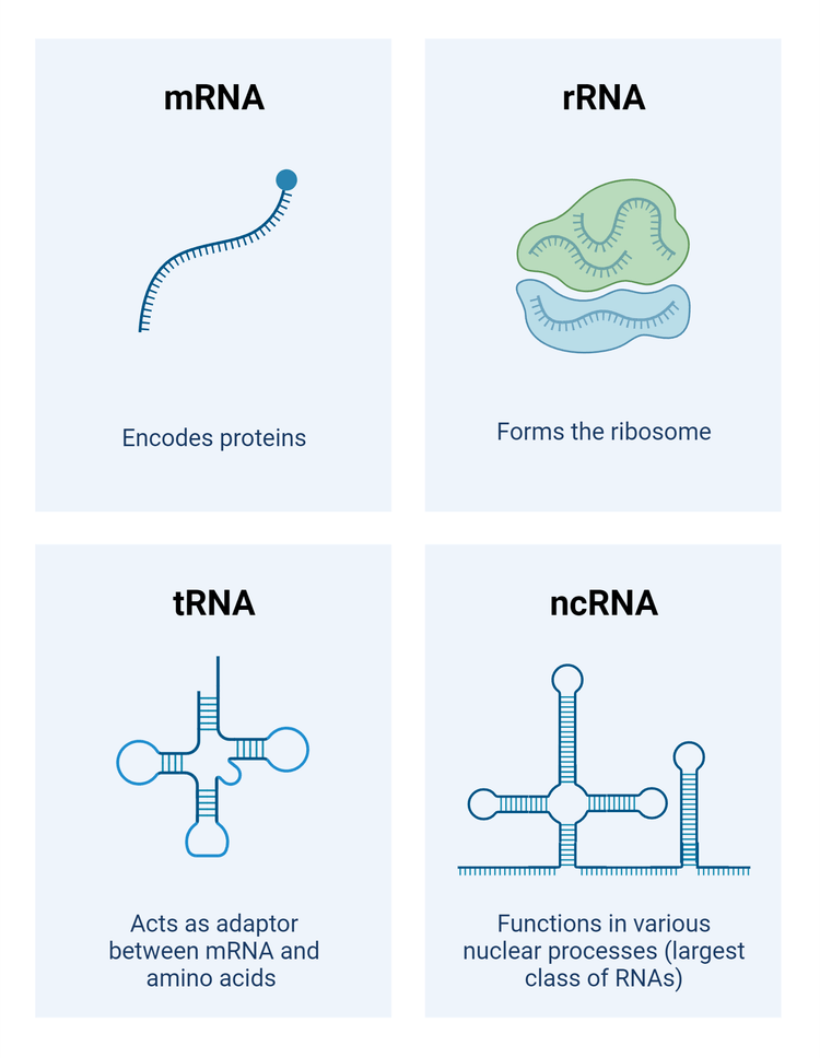
Examples of some common nucleic acid types, including mRNA, tRNA, rRNA, & ncRNA. Figure made in BioRender.
- Messenger RNA (mRNA), for example, carries genetic information copied from DNA to help with the creation of proteins. mRNAs are long, generally single-stranded, molecules that consist of nucleotides attached by phosphodiester bonds. mRNAs have a 7-methylguanosine cap on their 5' end that protects it from degradation, and a poly(A) tail on their 3' end that aids in stability during transport.
- Another type of RNA, transfer RNA (tRNA), helps translate mRNA into proteins. tRNAs exhibit a cloverleaf structure and are specific to each amino acid. This structure consists of a 3' acceptor site, a 5' terminal phosphate, and four “arms” or “loops” that are considered the business ends of the molecule.
- Ribosomal RNA (rRNA) is another variant of RNA that exists in a spherical shape. rRNA provides the structural framework for ribosomes which, essential to protein synthesis. Ribosomes contain a large and small ribosomal subunit with an exit (E), peptidyl (P), and acceptor (A) site.
- Alternatively, non-coding RNA (ncRNAs) are transcripts that do not encode proteins and cumulatively make up the biggest class of RNAs. ncRNAs are most easily divided into small (<200 nucleotides) and long ncRNAs (>200 nucleotides), that vary in structure and function within the cell.
Artificial Nucleic Acids
Today there exist many types of artificial nucleic acid analogues, also termed xenonucleic acids (XNAs), which are distinguished from naturally occurring nucleic acids due to alterations in their backbones and/or nucleobases. XNAs have great potential in biomedicine for use as potential therapeutic agents and/or fluorescent probes for medical research. XNAs mimic naturally occurring nucleic acids but have several advantages including facile chemical synthesis and resistance to nuclease-mediated cleavage. For example, peptide nucleic acid (PNA) is a non-charged oligonucleotide analogue that contains an N-aminoethyl glycine-based polyamide structure instead of a sugar backbone. In contrast to natural DNA, PNA binds in antiparallel and parallel orientations to complementary nucleic acids.
| Polyamide | Polyimide |
| Is a synthetic polymer made by the linkage of an amino group of one molecule, and a carboxylic acid group of another molecule | Is a very strong polymer made from imide monomers |
| Has recurring amide linkages | Has recurring imide linkages |
| Its monomers are diamines and dicarboxylic acids | Its monomers are either dianhydride and diisocyanate or dianhydride and diamine |
| Examples are nylon, Kevlar (synthetic polyamides) and wool and silk (natural polyamides) | Examples are Kapton, Apical, and Kaptrex |
Locked nucleic acids (LNA) are another form of synthetic oligonucleotides. LNAs have one or more nucleotides in which an extra methylene bridge can fix the ribose backbone into two separate conformations. LNA is particularly attractive for in vivo applications. In the literature, LNA has shown to have increased affinity over other RNA, can exhibit improved mismatch discrimination, has low toxicity, and has increased metabolic stability.
Currently, dozens of other XNAs exist with various conformations. Some other common XNAs include threose nucleic acid (TNA, with a simplified four-carbon threose backbone), glycol nucleic acid (GNA, based on a glycol monomer), and hexitol nucleic acid (HNA, that has a 1',5'-anhydrohexitol sugar).
Product Ordering Information
Table 3. Product ordering information for Gelite™ Safe DNA Gel Stain
| Product ▲ ▼ | Size ▲ ▼ | Cat No. ▲ ▼ |
| Gelite™ Safe DNA Gel Stain *10,000X Water Solution* | 100 µL | 17700 |
| Gelite™ Safe DNA Gel Stain *10,000X Water Solution* | 500 µL | 17701 |
| Gelite™ Safe DNA Gel Stain *10,000X Water Solution* | 1 mL | 17702 |
| Gelite™ Safe DNA Gel Stain *10,000X Water Solution* | 10 mL | 17703 |
| Gelite™ Safe DNA Gel Stain *10,000X DMSO Solution* | 100 µL | 17704 |
| Gelite™ Safe DNA Gel Stain *10,000X DMSO Solution* | 500 µL | 17705 |
| Gelite™ Safe DNA Gel Stain *10,000X DMSO Solution* | 1 mL | 17706 |
| Gelite™ Safe DNA Gel Stain *10,000X DMSO Solution* | 10 mL | 17707 |
| Gelite™ Safe DNA Gel Stain *GelRed Replacement, 10,000X in water* | 0.5 mL | 17708 |
| Gelite™ Safe DNA Gel Stain *GelRed Replacement, 10,000X in DMSO* | 0.5 mL | 17709 |
Table 4. Hoechst dyes for labeling DNA in fluorescence microscopy and flow cytometry.
| Product ▲ ▼ | Ex (nm) ▲ ▼ | Em (nm) ▲ ▼ | Unit Size ▲ ▼ | Cat. No. ▲ ▼ |
| Hoechst 33342 *Ultrapure Grade* | 352 | 454 | 100 mg | 17530 |
| Hoechst 33342 *Ultrapure Grade* | 352 | 454 | 1 g | 17533 |
| Hoechst 33342 *20 mM solution in water* | 352 | 454 | 5 mL | 17535 |
| Hoechst 33258 | 352 | 454 | 100 mg | 17520 |
| Hoechst 33258 | 352 | 454 | 1 g | 17523 |
| Hoechst 33258 *20 mM solution in water* | 352 | 454 | 5 mL | 17525 |
| Hoechst 34580 | 371 | 438 | 5 mg | 17537 |
| Hoechst 34580 *20 mM solution in water* | 371 | 438 | 100 µL | 17538 |
| Nuclear Yellow (Hoechst 33258) | 372 | 504 | 5 mL | 17539 |
Table 5. RNA quantification and PCR reagents
| Product Name ▲ ▼ | Ex (nm) ▲ ▼ | Em (nm) ▲ ▼ | Unit Size ▲ ▼ | Cat No. ▲ ▼ |
| StrandBrite™ Green Fluorimetric RNA Quantitation Kit *Optimized for Microplate Readers* | 490 nm | 545 nm | 1000 Tests | 17655 |
| StrandBrite™ Green Fluorimetric RNA Quantitation Kit | 490 nm | 540 nm | 100 Tests | 17656 |
| StrandBrite™ Green Fluorimetric RNA Quantitation Kit *High Selectivity* | 490 nm | 540 nm | 100 Tests | 17657 |
| StrandBrite™ Green RNA Quantifying Reagent | 490 nm | 525 nm | 1 mL | 17610 |
| StrandBrite™ Green RNA Quantifying Reagent | 490 nm | 525 nm | 10 mL | 17611 |
| Portelite™ Fluorimetric RNA Quantitation Kit | 490 nm | 525 nm | 100 Tests | 17658 |
| Portelite™ Fluorimetric RNA Quantitation Kit | 490 nm | 525 nm | 500 Tests | 17659 |
| Cyber Green™ [Equivalent to SYBR® Green] *20X Aqueous PCR Solution* | 498 nm | 522 nm | 5 x 1 mL Tests | 17591 |
| Cyber Green™ [Equivalent to SYBR® Green] *20X Aqueous PCR Solution* | 498 nm | 522 nm | 1 mL | 17592 |
| Cyber Green™ Nucleic Acid Gel Stain [Equivalent to SYBR® Green] | 498 nm | 522 nm | 100 µL | 17604 |
Table 6. DNA Sequencing Building Blocks
| Cat# ▲ ▼ | Product Name ▲ ▼ | Unit Size ▲ ▼ |
| 300 | 5-dR6G [5-Carboxy-4,7-dichlororhodamine 6G] | 25 mg |
| 301 | 6-dR6G [6-Carboxy-4,7-dichlororhodamine 6G] | 25 mg |
| 302 | 5-dR6G, succinimidyl ester | 5 mg |
| 305 | 5-dTMR [5-Carboxy-4,7-dichlorortetramethylrhodamine] | 25 mg |
| 306 | 6-dTMR [6-Carboxy-4,7-dichlorortetramethylrhodamine] | 25 mg |
| 307 | 5-dTMR, succinimidyl ester | 5 mg |
| 310 | 5-dROX [5-Carboxy-4,7-dichloror-X-hodamine] | 25 mg |
| 311 | 6-dROX [6-Carboxy-4,7-dichloror-X-hodamine] | 25 mg |
| 312 | 5-dROX, succinimidyl ester | 5 mg |
| 315 | 5-dR110 [5-Carboxy-4,7-dichlororhodamine 110] | 25 mg |
References
Deoxyribonucleic acid (DNA)
Understanding biochemistry: structure and function of nucleic acids
Biochemistry, RNA Structure
Antisense: Medicinal Chemistry
Chapter 13 - Zebrafish as a Research Organism: Danio rerio in Biomedical Research
Locked nucleic acid oligonucleotides: the next generation of antisense agents?
Acyclic (S)-glycol nucleic acid (S-GNA) modification of siRNAs improves the safety of RNAi therapeutics while maintaining potency