ReadiLink™ Rapid XFD750 Antibody Labeling Kit *Production Scale*
| Price | |
| Catalog Number | |
| Unit Size | |
| Quantity |
| Telephone | 1-800-990-8053 |
| Fax | 1-800-609-2943 |
| sales@aatbio.com | |
| International | See distributors |
| Bulk request | Inquire |
| Custom size | Inquire |
| Shipping | Standard overnight for United States, inquire for international |
| Correction Factor (260 nm) | 0.00 |
| Correction Factor (280 nm) | 0.04 |
| Extinction coefficient (cm -1 M -1) | 240000 |
| Excitation (nm) | 752 |
| Emission (nm) | 776 |
| Quantum yield | 0.121 |
| H-phrase | H303, H313, H333 |
| Hazard symbol | XN |
| Intended use | Research Use Only (RUO) |
| R-phrase | R20, R21, R22 |
| UNSPSC | 12171501 |
| ReadiLink™ Rapid iFluor® 750 Antibody Labeling Kit *Microscale Optimized for Labeling 50 μg Antibody Per Reaction* |
| ReadiLink™ Rapid iFluor® 750 Antibody Labeling Kit *Production Scale* |
| ReadiLink™ Rapid XFD750 Antibody Labeling Kit *XFD750 Same Structure to Alexa Fluor™ 750* |
| Overview |
Correction Factor (260 nm) 0.00 | Correction Factor (280 nm) 0.04 | Extinction coefficient (cm -1 M -1) 240000 | Excitation (nm) 752 | Emission (nm) 776 | Quantum yield 0.121 |
Components
Example protocol
AT A GLANCE
1.0 mg Antibody (MW ~150 kDa)
Antibody concentration: 2.0 mg/mL
Antibody volume: 500 µL
SAMPLE EXPERIMENTAL PROTOCOL
Before opening the vials, warm all components and briefly centrifuge. Immediately prepare necessary solutions before starting conjugation. This protocol is a recommendation.
Warm up a vial of reactive dye (Component A) to room temperature.
Note: Each vial of reactive dye contains an optimized amount of dye to label 1 mg of IgG (MW ~150 kDa) at 2 mg/mL in PBS, the kit can also be used to label other proteins (>10 kDa).
Add 10 µL of DMSO (Component D) to the vial of reactive dye (Component A), mix well.
Prepare a 500 µL antibody solution in PBS with a concentration of 2 mg/mL.
Note: The protein should be dissolved in 1X phosphate buffered saline (PBS), pH 7.2 - 7.4. If the protein is dissolved in buffers containing primary amines, like Tris and/or glycine, it must be dialyzed against 1X PBS, pH 7.2 - 7.4, or use Amicon Ultra0.5, Ultracel-10 Membrane, 10 kDa (Cat No. UFC501008 from Millipore) to remove free amines or ammonium salts (such as ammonium sulfate and ammonium acetate) that are widely used for protein precipitation.
Note: Impure antibodies or antibodies stabilized with bovine serum albumin (BSA) or gelatin will not be labeled well.
Add 25 µL of Reaction Buffer (Component B) to the antibody solution.
Transfer the reconstituted dye solution into the vial of antibody solution, and pipette several times to mix well.
Rotate the reaction mixture for 1 hour at room temperature.
Twist off the bottom closure of the desalting column (Component D), and loosen the cap. Place the column in a collection tube.
Centrifuge the column at 1,000 g for 2 minutes to remove the storage solution.
Remove the cap and slowly add 1 mL of PBS to the column. Centrifuge at 1,000 g for 2 minutes and remove the buffer. Repeat this step 3 additional times, discarding the buffer from the collection tube each time.
Place the column in a new collection tube, and gently apply the sample into the center of the compact resin bed.
Centrifuge the column at 1,000 g for 2 minutes to collect the sample.
The following formula can be used to calculate the antibody concentration:
(A280 - CF280 x Adye) / 1.4
The following formula can be used to calculate the degree of labeling:
DOL = (Adye / Ecdye) / (A280 - CF280 x Adye) / 210,000)
Where:
- 210,000 is the molar extinction coefficient (Ec) in cm-1M-1 of IgG at 280 nm.
- CF280 is the correction factor for the effect of the fluorophore on absorbance at 280 nm.
- Adye is the absorbance at maximum (λmax) for the respective dye.
Table 1. Properties of Labeling Dyes found in the ReadiLink™ Rapid Antibody Labeling Kits.
Cat# | Dye | Mol. Wt. | Ec (cm-1M-1) | CF280 | Target DOL |
5700 | iFluor® 350 | 749.85 | 20,000 | 0.23 | 5-10 |
5702 | iFluor® 488 | 945.07 | 75,000 | 0.21 | 4-8 |
5705 | iFluor® 555 | 914.06 | 90,000 | 0.16 | 4-7 |
5710 | iFluor® 594 | 1160.42 | 18,000 | 0.04 | 3-6 |
5713 | iFluor® 647 | 1274.66 | 250,000 | 0.03 | 3-7 |
5718 | iFluor® 750 | 1416.83 | 250,000 | 0.039 | 2-6 |
5720 | FITC | 620.52 | 75,000 | 0.183 | 3-6 |
5722 | Cy3 | 829.03 | 150,000 | 0.073 | 1-3 |
5725 | Cy5 | 855.07 | 250,000 | 0.03 | 2-4 |
5727 | Cy7 | 881.11 | 250,000 | 0.036 | 2-4 |
5730 | XFD488 | 643.4 | 71,000 | 0.11 | 4-8 |
5733 | XFD555 | 1250 | 150,000 | 0.08 | 4-7 |
5736 | XFD594 | 819.85 | 90,000 | 0.56 | 3-6 |
5740 | XFD647 | 1259.66 | 240,000 | 0.03 | 3-7 |
5745 | XFD750 | 1300 | 240,000 | 0.04 | 2-5 |
Spectrum
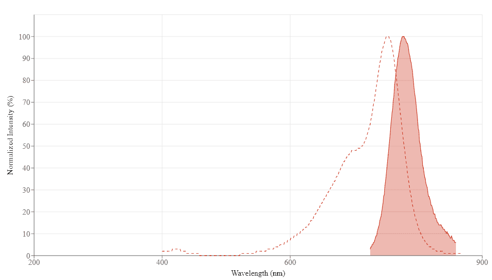
Spectral properties
| Correction Factor (260 nm) | 0.00 |
| Correction Factor (280 nm) | 0.04 |
| Extinction coefficient (cm -1 M -1) | 240000 |
| Excitation (nm) | 752 |
| Emission (nm) | 776 |
| Quantum yield | 0.121 |
Product Family
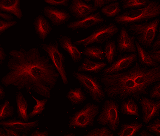
Images

References
Authors: Yan, Pengyu and Chen, Dan and Yan, Xutao and Yan, Xiaoting and Wang, Yingpeng and Liu, Chao and Yang, Xiaofeng
Journal: Frontiers in oncology (2022): 825476
Authors: Yang, Yongjun and Yan, Xiaoting and Li, Jiawei and Liu, Chao and Yang, Xiaofeng
Journal: Molecular therapy oncolytics (2022): 319-330
Authors: Ovejero Paredes, Karina and Díaz-García, Diana and García-Almodóvar, Victoria and Lozano Chamizo, Laura and Marciello, Marzia and Díaz-Sánchez, Miguel and Prashar, Sanjiv and Gómez-Ruiz, Santiago and Filice, Marco
Journal: Cancers (2020)
Authors: Kantamneni, Harini and Barkund, Shravani and Donzanti, Michael and Martin, Daniel and Zhao, Xinyu and He, Shuqing and Riman, Richard E and Tan, Mei Chee and Pierce, Mark C and Roth, Charles M and Ganapathy, Vidya and Moghe, Prabhas V
Journal: BMC cancer (2020): 1082
Authors: Rudkouskaya, Alena and Sinsuebphon, Nattawut and Ochoa, Marien and Chen, Sez-Jade and Mazurkiewicz, Joseph E and Intes, Xavier and Barroso, Margarida
Journal: Theranostics (2020): 10309-10325
Authors: Lei, Li and Li, Min and Wu, Sufen and Xu, Zhiai and Geng, Ping and Tian, Yang and Fu, Ying and Zhang, Wen
Journal: Analytical chemistry (2020): 5838-5845
Authors: Panagia, Marcello and Yang, Jing and Gale, Eric and Wang, Huan and Luptak, Ivan and Chen, Howard H and Patel, Dakshesh and Croteau, Dominique and Pimentel, David Richard and Bachschmid, Markus Michael and Colucci, Wilson S and Ran, Chongzhao and Sosnovik, David E
Journal: Scientific reports (2020): 11209
Authors: Kleinmanns, Katrin and Fosse, Vibeke and Davidson, Ben and de Jalón, Elvira García and Tenstad, Olav and Bjørge, Line and McCormack, Emmet
Journal: EBioMedicine (2020): 102783
Authors: Yang, Yongjun and Yang, Xiaofeng and Liu, Chao and Li, Jiawei
Journal: World journal of urology (2020)
Authors: Byrd, Brook K and Folaron, Margaret R and Leonor, Joseph P and Strawbridge, Rendall R and Cao, Xu and Bruza, Petr and Davis, Scott C
Journal: Journal of biomedical optics (2019): 1-5
Application notes
A New Protein Crosslinking Method for Labeling and Modifying Antibodies
A Novel Fluorescent Probe for Imaging and Detecting Hydroxyl Radical in Living Cells
A Novel NO Wash Probeniceid-Free Calcium Assay for Functional Analysis of GPCR and Calcium Channel Targets
Abbreviation of Common Chemical Compounds Related to Peptides
FAQ
Are there any alternatives to BrdU (Bromodeoxyuridine)?
Are there any alternatives to Cy5?
Are there any alternatives to indocyanine green (ICG)?
Are there any calcium indicators that don't require probenecid (PBC)?