A Meta-Analysis of Common Calcium Indicators
Calcium is one of the most prevalent ions involved in eukaryote physiology, taking part in vital processes from bone development to signal transduction, and disorders in intracellular calcium regulation can lead to severe neurological and structural complications. Because of its importance, calcium is a relatively large topic of interest to study at the cellular level. In order to visualize and quantify intracellular calcium, researchers have employed fluorescent calcium indicators (or dyes) which can chelate free floating calcium ions to create a quantitative fluorogenic response. When paired with fluorescence microscopes, microplate readers, or flow cytometers, calcium indicators are useful for imaging and monitoring intracellular calcium concentrations.
Fluorescent calcium indicators can be either genetically or chemically engineered. Genetic indicators involve transfection or expression of a variant of GFP in a live cell or animal, while chemical indicators consist of small molecules based on BAPTA, which has high selectivity for calcium ions. Out of these two major groups of calcium indicators, chemical indicators are mainly favored because of their commercial availability, convenience, and flexibility in experimental design.
Among the current selection of chemical indicators are the fairly popular single-wavelength calcium indicators, which will fluoresce in only one color whether or not they are bound to calcium. Traditional green fluorescent calcium indicators include Fluo-3, and its improved version, Fluo-4. Fluo-4 was developed by Gee and colleagues in 2000. It is essentially a non-fluorescent substrate on its own, but once it is bound to calcium its fluorescence increases by 100-fold. Its imaging has allowed researchers to uncover many interactions involved in calcium signaling, and more recently, it has been widely incorporated into cell-based HTS assays for drug discovery. Fluo-4 is an analog of Fluo-3 with two chlorine atoms replaced by fluorine atoms, resulting in an excitation peak shift 12nm closer to the 488nm of an argon laser or another source in conjunction with the standard fluorescein filter set.
Despite its slight structural modifications from Fluo-3, Fluo-4 demonstrates a large improvement in producing a brighter and more sensitive signal, making Fluo-4 ideal for confocal microscopy, flow cytometry, and microplate screening applications. Fluo-4's cell-permeable variant, Fluo-4 AM, is a popular green fluorescent calcium indicator that does not require a quench for background noise.
Even with its 100-fold increase in fluorescence, Fluo-4 AM is not without its problems in a few areas. One common issue encountered is a low signal-to-background ratio, due to cell leakage. To address this issue, most indicators from the Fluo series will be paired with a reagent that either prevents the fluorescent reaction from leaving the cell or dissolves the leakage to reduce background signal. Examples of these reagents include probenecid or sulfinpyrazone which inhibit anion transporters in the cells membranes to prevent any passage of calcium. A common compatible surfactant is Pluronic® polyols which help dissolve the dyes in aqueous solution. However, applications of these substances subject the cellular environment to more invasive procedures and may affect overall cell integrity or activity of calcium. To avoid the use of extra reagents, one alternative available is Cal-520 AM. The green dye Cal-520 has been optimized to address the issue of dye loss with its improved intracellular retention, such that no extra reagents are required to keep calcium contained or background suppressed. Cal-520 enables higher sensitivity and signal-to-background ratios at a spectrum similar to that of Fluo-3 and Fluo-4.
Besides dye loss, an additional drawback from using Fluo-4 is maintaining the harsh cell loading conditions that are required of its use. An alternative green dye, Fluo-8, makes it possible to load cells at room temperature instead of the usual 37 degrees Celsius that is needed for Fluo-3 and Fluo-4. Since Fluo-8's performance is not as temperature-dependent, it's the perfect candidate for use in HTS applications. Fluo-8 is also prepared at different dissociation constants (higher and lower affinity) and packaging sizes, including an HTS-ready size. And last but not least, Fluo-8 emits a signal that is two times stronger than that of Fluo-4. Like Cal-520, Fluo-8 shares very similar excitation and emission peaks with Fluo-4, while being a more accessible, versatile option to conducting green fluorescence imaging.
Another troublesome aspect of chemical calcium indicators is their tendency to compartmentalize within a cell. One dye that exhibits this phenomenon is Rhod-2 AM, a popular red calcium indicator. Its net positive charge encourages sequestration into mitochondria in many types of cells. This localization of Rhod-2 in the mitochondria occurs shortly after its introduction into the cell, resulting in a limited fluorescent signal within live cells. Cal-590 was designed to remain localized in a cell's cytosol, instead of its mitochondria, vacuole, or other organelles, while retaining Rhod-2's spectral wavelength. Cal-590 is good for comprehensive overall cellular imaging for red dye imaging. There are also dextran forms of calcium indicators available for an even more effective measure in both compartmentalization and intracellular dye retention. Addition of the large molecule dextran to a dye molecule allows for high solubility while maintaining low toxicity, which makes it perfect as a preventative for compartmentalization or leakage when conjugated to dyes. The only shortcoming that arises from choosing to use dextran conjugates it that it has to be injected into the cell, either by patch pipette or microinjection.
Beyond enhanced fluorescence and improved cytosolic retention, The Cal-series of dyes (Cal-520, Cal-590, and Cal-630) possess spectral qualities for multiplexing or multicolor detection. Cal-520 holds its excitation maxima at 492, which is closer to an argon laser's 488 nm than the excitation maxima for other green dyes. This proximity to 488 nm makes Cal-520 readily compatible with any fluorescence instrumentation platforms. With excitation maxima at 573 and 608 nm, and emissions at 588 and 626 nm, respectively, the long wavelengths of the red Cal-590 and Cal-630 make them compatible with green fluorescent protein (GFP) cell lines, as well as FITC and AlexaFluor 488 labeled antibodies.
Fluorescent calcium indicators can be either genetically or chemically engineered. Genetic indicators involve transfection or expression of a variant of GFP in a live cell or animal, while chemical indicators consist of small molecules based on BAPTA, which has high selectivity for calcium ions. Out of these two major groups of calcium indicators, chemical indicators are mainly favored because of their commercial availability, convenience, and flexibility in experimental design.

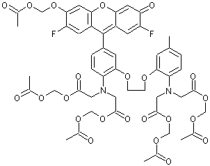
Chemical Structure of Fluo-4
Despite its slight structural modifications from Fluo-3, Fluo-4 demonstrates a large improvement in producing a brighter and more sensitive signal, making Fluo-4 ideal for confocal microscopy, flow cytometry, and microplate screening applications. Fluo-4's cell-permeable variant, Fluo-4 AM, is a popular green fluorescent calcium indicator that does not require a quench for background noise.
Even with its 100-fold increase in fluorescence, Fluo-4 AM is not without its problems in a few areas. One common issue encountered is a low signal-to-background ratio, due to cell leakage. To address this issue, most indicators from the Fluo series will be paired with a reagent that either prevents the fluorescent reaction from leaving the cell or dissolves the leakage to reduce background signal. Examples of these reagents include probenecid or sulfinpyrazone which inhibit anion transporters in the cells membranes to prevent any passage of calcium. A common compatible surfactant is Pluronic® polyols which help dissolve the dyes in aqueous solution. However, applications of these substances subject the cellular environment to more invasive procedures and may affect overall cell integrity or activity of calcium. To avoid the use of extra reagents, one alternative available is Cal-520 AM. The green dye Cal-520 has been optimized to address the issue of dye loss with its improved intracellular retention, such that no extra reagents are required to keep calcium contained or background suppressed. Cal-520 enables higher sensitivity and signal-to-background ratios at a spectrum similar to that of Fluo-3 and Fluo-4.
Besides dye loss, an additional drawback from using Fluo-4 is maintaining the harsh cell loading conditions that are required of its use. An alternative green dye, Fluo-8, makes it possible to load cells at room temperature instead of the usual 37 degrees Celsius that is needed for Fluo-3 and Fluo-4. Since Fluo-8's performance is not as temperature-dependent, it's the perfect candidate for use in HTS applications. Fluo-8 is also prepared at different dissociation constants (higher and lower affinity) and packaging sizes, including an HTS-ready size. And last but not least, Fluo-8 emits a signal that is two times stronger than that of Fluo-4. Like Cal-520, Fluo-8 shares very similar excitation and emission peaks with Fluo-4, while being a more accessible, versatile option to conducting green fluorescence imaging.
Another troublesome aspect of chemical calcium indicators is their tendency to compartmentalize within a cell. One dye that exhibits this phenomenon is Rhod-2 AM, a popular red calcium indicator. Its net positive charge encourages sequestration into mitochondria in many types of cells. This localization of Rhod-2 in the mitochondria occurs shortly after its introduction into the cell, resulting in a limited fluorescent signal within live cells. Cal-590 was designed to remain localized in a cell's cytosol, instead of its mitochondria, vacuole, or other organelles, while retaining Rhod-2's spectral wavelength. Cal-590 is good for comprehensive overall cellular imaging for red dye imaging. There are also dextran forms of calcium indicators available for an even more effective measure in both compartmentalization and intracellular dye retention. Addition of the large molecule dextran to a dye molecule allows for high solubility while maintaining low toxicity, which makes it perfect as a preventative for compartmentalization or leakage when conjugated to dyes. The only shortcoming that arises from choosing to use dextran conjugates it that it has to be injected into the cell, either by patch pipette or microinjection.
Beyond enhanced fluorescence and improved cytosolic retention, The Cal-series of dyes (Cal-520, Cal-590, and Cal-630) possess spectral qualities for multiplexing or multicolor detection. Cal-520 holds its excitation maxima at 492, which is closer to an argon laser's 488 nm than the excitation maxima for other green dyes. This proximity to 488 nm makes Cal-520 readily compatible with any fluorescence instrumentation platforms. With excitation maxima at 573 and 608 nm, and emissions at 588 and 626 nm, respectively, the long wavelengths of the red Cal-590 and Cal-630 make them compatible with green fluorescent protein (GFP) cell lines, as well as FITC and AlexaFluor 488 labeled antibodies.
Original created on September 15, 2016, last updated on September 15, 2016
Tagged under: