ReadiLink™ Rapid iFluor® 594 Antibody Labeling Kit *Production Scale*
| Price | |
| Catalog Number | |
| Unit Size | |
| Quantity |
| Telephone | 1-800-990-8053 |
| Fax | 1-800-609-2943 |
| sales@aatbio.com | |
| International | See distributors |
| Bulk request | Inquire |
| Custom size | Inquire |
| Shipping | Standard overnight for United States, inquire for international |
| Solvent | DMSO |
| Absorbance (nm) | 587 |
| Correction Factor (260 nm) | 0.05 |
| Correction Factor (280 nm) | 0.04 |
| Extinction coefficient (cm -1 M -1) | 2000001 |
| Excitation (nm) | 587 |
| Emission (nm) | 603 |
| Quantum yield | 0.531 |
| H-phrase | H303, H313, H333 |
| Hazard symbol | XN |
| Intended use | Research Use Only (RUO) |
| R-phrase | R20, R21, R22 |
| Storage | Freeze (< -15 °C); Minimize light exposure |
| UNSPSC | 12171501 |
| ReadiLink™ Rapid iFluor® 594 Antibody Labeling Kit *Microscale Optimized for Labeling 50 μg Antibody Per Reaction* |
| Overview |
Absorbance (nm) 587 | Correction Factor (260 nm) 0.05 | Correction Factor (280 nm) 0.04 | Extinction coefficient (cm -1 M -1) 2000001 | Excitation (nm) 587 | Emission (nm) 603 | Quantum yield 0.531 |
Components
Example protocol
AT A GLANCE
1.0 mg Antibody (MW ~150 kDa)
Antibody concentration: 2.0 mg/mL
Antibody volume: 500 µL
SAMPLE EXPERIMENTAL PROTOCOL
Before opening the vials, warm all components and briefly centrifuge. Immediately prepare necessary solutions before starting conjugation. This protocol is a recommendation.
Warm up a vial of reactive dye (Component A) to room temperature.
Note: Each vial of reactive dye contains an optimized amount of dye to label 1 mg of IgG (MW ~150 kDa) at 2 mg/mL in PBS, the kit can also be used to label other proteins (>10 kDa).
Add 10 µL of DMSO (Component D) to the vial of reactive dye (Component A), mix well.
Prepare a 500 µL antibody solution in PBS with a concentration of 2 mg/mL.
Note: The protein should be dissolved in 1X phosphate buffered saline (PBS), pH 7.2 - 7.4. If the protein is dissolved in buffers containing primary amines, like Tris and/or glycine, it must be dialyzed against 1X PBS, pH 7.2 - 7.4, or use Amicon Ultra0.5, Ultracel-10 Membrane, 10 kDa (Cat No. UFC501008 from Millipore) to remove free amines or ammonium salts (such as ammonium sulfate and ammonium acetate) that are widely used for protein precipitation.
Note: Impure antibodies or antibodies stabilized with bovine serum albumin (BSA) or gelatin will not be labeled well.
Add 25 µL of Reaction Buffer (Component B) to the antibody solution.
Transfer the reconstituted dye solution into the vial of antibody solution, and pipette several times to mix well.
Rotate the reaction mixture for 1 hour at room temperature.
Twist off the bottom closure of the desalting column (Component D), and loosen the cap. Place the column in a collection tube.
Centrifuge the column at 1,000 g for 2 minutes to remove the storage solution.
Remove the cap and slowly add 1 mL of PBS to the column. Centrifuge at 1,000 g for 2 minutes and remove the buffer. Repeat this step 3 additional times, discarding the buffer from the collection tube each time.
Place the column in a new collection tube, and gently apply the sample into the center of the compact resin bed.
Centrifuge the column at 1,000 g for 2 minutes to collect the sample.
The following formula can be used to calculate the antibody concentration:
(A280 - CF280 x Adye) / 1.4
The following formula can be used to calculate the degree of labeling:
DOL = (Adye / Ecdye) / (A280 - CF280 x Adye) / 210,000)
Where:
- 210,000 is the molar extinction coefficient (Ec) in cm-1M-1 of IgG at 280 nm.
- CF280 is the correction factor for the effect of the fluorophore on absorbance at 280 nm.
- Adye is the absorbance at maximum (λmax) for the respective dye.
Table 1. Properties of Labeling Dyes found in the ReadiLink™ Rapid Antibody Labeling Kits.
Cat# | Dye | Mol. Wt. | Ec (cm-1M-1) | CF280 | Target DOL |
5700 | iFluor® 350 | 749.85 | 20,000 | 0.23 | 5-10 |
5702 | iFluor® 488 | 945.07 | 75,000 | 0.21 | 4-8 |
5705 | iFluor® 555 | 914.06 | 90,000 | 0.16 | 4-7 |
5710 | iFluor® 594 | 1160.42 | 18,000 | 0.04 | 3-6 |
5713 | iFluor® 647 | 1274.66 | 250,000 | 0.03 | 3-7 |
5718 | iFluor® 750 | 1416.83 | 250,000 | 0.039 | 2-6 |
5720 | FITC | 620.52 | 75,000 | 0.183 | 3-6 |
5722 | Cy3 | 829.03 | 150,000 | 0.073 | 1-3 |
5725 | Cy5 | 855.07 | 250,000 | 0.03 | 2-4 |
5727 | Cy7 | 881.11 | 250,000 | 0.036 | 2-4 |
5730 | XFD488 | 643.4 | 71,000 | 0.11 | 4-8 |
5733 | XFD555 | 1250 | 150,000 | 0.08 | 4-7 |
5736 | XFD594 | 819.85 | 90,000 | 0.56 | 3-6 |
5740 | XFD647 | 1259.66 | 240,000 | 0.03 | 3-7 |
5745 | XFD750 | 1300 | 240,000 | 0.04 | 2-5 |
Spectrum
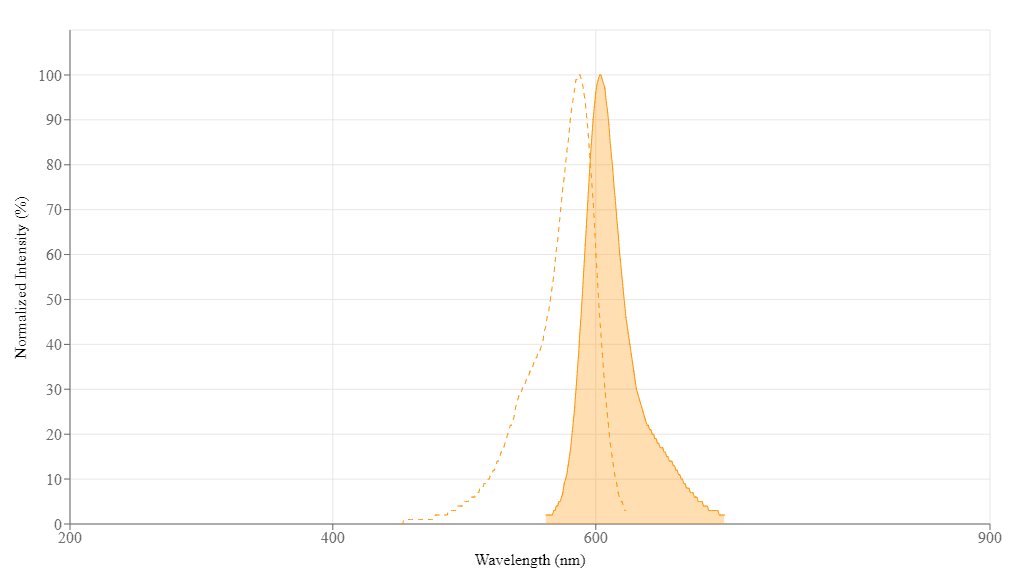
Spectral properties
| Absorbance (nm) | 587 |
| Correction Factor (260 nm) | 0.05 |
| Correction Factor (280 nm) | 0.04 |
| Extinction coefficient (cm -1 M -1) | 2000001 |
| Excitation (nm) | 587 |
| Emission (nm) | 603 |
| Quantum yield | 0.531 |
Product Family
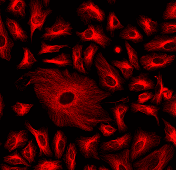
Images

References
Authors: Ferraro, G and Gigante, Y and Pitea, M and Mautone, L and Ruocco, G and Di Angelantonio, S and Leonetti, M
Journal: Scientific reports (2023): 10983
Authors: Qin, Mengyuan and Huang, Jie and Zhong, Jincheng and Zhang, Yingxian and Tong, Shen and Cheng, Hui and Deng, Xiangquan and Zheng, Lei and Zhang, Wanjian and Qiu, Ping and Wang, Ke
Journal: Journal of biophotonics (2023): e202200365
Authors: Zhong, Chuanmei and Ju, Gongchenhao and Yang, Sufang and Zhao, Xiangpei and Chen, Jixiang and Li, Ning
Journal: Gynecologic and obstetric investigation (2023): 197-213
Authors: Chung, Hye Yoon and Kim, Min Ju and Kim, Seung Woo and Oh, Jeeyoung and Shin, Ha Young
Journal: Journal of clinical neurology (Seoul, Korea) (2023): 60-66
Authors: Spahr, Zachary R and Economides, John R and Horton, Jonathan C
Journal: Journal of neuro-ophthalmology : the official journal of the North American Neuro-Ophthalmology Society (2022): e596-e597
Authors: Leibrand, Crystal R and Paris, Jason J and Jones, Austin M and Ohene-Nyako, Michael and Rademeyer, Kara M and Nass, Sara R and Kim, Woong-Ki and Knapp, Pamela E and Hauser, Kurt F and McRae, MaryPeace
Journal: Neuroscience letters (2022): 136852
Authors: Zhao, Yan and Liu, Shuo and Shi, Zhishang and Zhu, Hangqi and Li, Mingchun and Yu, Qilin
Journal: Nano research (2022): 6243-6255
Authors: Xu, Ling-Ling and Yan, Yao and Yuan, Yu-Min and Li, Ying and Jiang, Jun and Zhang, Li-Cai
Journal: Journal of pain research (2022): 3931-3939
Authors: Alfirdous, Rayyan A and Alquiria, Theeb A and Jacinto, Rogerio C and Martinho, Frederico C
Journal: International endodontic journal (2022): 1081-1090
Authors: Atis, Muge and Akcan, Uğur and Altunsu, Deniz and Ayvaz, Ecem and Uğur Yılmaz, Canan and Sarıkaya, Deniz and Temizyürek, Arzu and Ahıshalı, Bülent and Girouard, Hélène and Kaya, Mehmet
Journal: Brain research (2022): 148071
Application notes
A Novel Fluorescent Probe for Imaging and Detecting Hydroxyl Radical in Living Cells
A Novel NO Wash Probeniceid-Free Calcium Assay for Functional Analysis of GPCR and Calcium Channel Targets
Biotin Labeling Molecules and Their Biological Applications
Buccutite™ Bioconjugation Technology
FAQ
What are the differences between calcium ion indicators: Cal 520, Cal 520FF, and Cal 520N?
How do I make an AM ester stock solution?
Can we fix cells with glutaraldehyde and then stain with fluorescent phalloidin?
What is the difference between FluoroQuest Anti-fading Kit I and FluoroQuest Anti-fading Kit II?