Multiplexing Cell Proliferation and Cytotoxicity Assays Using Calcein Red and CytoCalceins
by Chunmei Wei, Haitao Guo, Jinfang Liao, Qinglin Meng, Xing Han, Zhenjun Diwu, Qin Zhao
Calcein AM is one of the most popular fluorescent probes used for labeling and monitoring cellular functions of live cells. However, the limited color selection of Calcein AM and its complete spectral overlap with GFP makes this valuable reagent impossible to be used in the multicolor analysis of live cells. Calcein Red™, Calcein Orange™ and CytoCalcein™ have been developed for multiplexing analysis of cellular functions. The non-fluorescent Calcein Red™, Calcein Orange™ and CytoCalcein™ become strongly fluorescent upon entering live cells. Calcein Red™, Calcein Orange™ and CytoCalcein™ are hydrophobic compounds that easily permeates intact live cells and generates their respective cell-retained fluorophores upon intracellular esterase-induced hydrolysis. The cellular esterase activity is proportional to the number of viable cells, and therefore directly related to the fluorescence intensity of Calcein Red™, Calcein Orange™ and CytoCalcein™. Using Calcein Orange™ and CytoCalcein™ cell proliferation and cell cytotoxicity were monitored in HEK, CPA, and Jurkat cells. The consistent results were obtained with flow cytometry, fluorescence plate reader and florescence microscopy. Calcein Orange™ and CytoCalcein™ have minimal cytotoxicity even after 2 weeks dye-loading in Jurkat cells while all the cells are apoptotic/necrotic after 3 days dye-loading with the same concentration of Calcein AM.


CytoCalcein™, Calcein Orange™ and Calcein Red™ are novel fluorescent probes that can be used with Calcein AM or other green fluorescent dyes for multicolor analysis. They have the following benefits and features:
Original created on January 16, 2010, last updated on January 16, 2010
Tagged under:
Introduction
Calcein AM is one of the most popular fluorescent probes used for labeling and monitoring cellular functions of live cells. However, the limited color selection of Calcein AM and its complete spectral overlap with GFP makes this valuable reagent impossible to be used in the multicolor analysis of live cells. Calcein Red™, Calcein Orange™ and CytoCalcein™ have been developed for multiplexing analysis of cellular functions. The non-fluorescent Calcein Red™, Calcein Orange™ and CytoCalcein™ become strongly fluorescent upon entering live cells. Calcein Red™, Calcein Orange™ and CytoCalcein™ are hydrophobic compounds that easily permeates intact live cells and generates their respective cell-retained fluorophores upon intracellular esterase-induced hydrolysis. The cellular esterase activity is proportional to the number of viable cells, and therefore directly related to the fluorescence intensity of Calcein Red™, Calcein Orange™ and CytoCalcein™. Using Calcein Orange™ and CytoCalcein™ cell proliferation and cell cytotoxicity were monitored in HEK, CPA, and Jurkat cells. The consistent results were obtained with flow cytometry, fluorescence plate reader and florescence microscopy. Calcein Orange™ and CytoCalcein™ have minimal cytotoxicity even after 2 weeks dye-loading in Jurkat cells while all the cells are apoptotic/necrotic after 3 days dye-loading with the same concentration of Calcein AM.
Materials and Methods
- CHO-M1, CPA, and Jurkat cells were plated in 96-well black wall/clear bottom costar plate at 37oC, 5% CO2 incubator for overnight.
- Dye-loading: After overnight incubation, culture medium was removed. 2-5 µM of Calcein, AM (Cat # 22004), Calcein Orange™ (Cat# 22009), Calcein Red™ (Cat# 22010), Calcein blue, AM (Cat # 22017), CytoCalcein™ Violet 450 (Cat# 22012), and CytoCalcein™ Violet 450 (Cat# 22012), in 100 µL of 1X HBSS with 20 mM HEPES was added into each well. The cells were dye-loaded at 37°C for 15 to 30 min. Cells were washed twice with buffer after dye loading.
- Run the experiments on a Fluorescence microscopy (Olympus IX71), BMG NOVOstar microplate reader or a FACSCalibur (Becton Dickinson, San Jose, CA).
Results
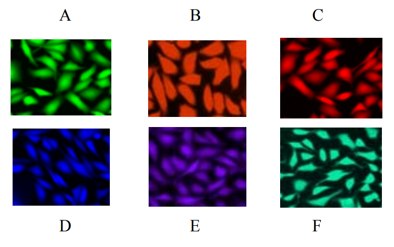
Image of CPA cells in a 96-well Costar black wall/clear bottom plate stained with different Calcein's. A: Calcein, AM (FITC filter), B: Calcein Orange™ (TRITC filter), C: Calcein Red™ (Cy5 filter), D: Calcein blue, AM (DAPI filter), E: CytoCalcein™ Violet 450 (Ex/Em = 405/450 nm, 405 Violet filter), F: CytoCalcein™ Violet 500 (Ex/Em = 405/500 nm, 405 Violet filter).

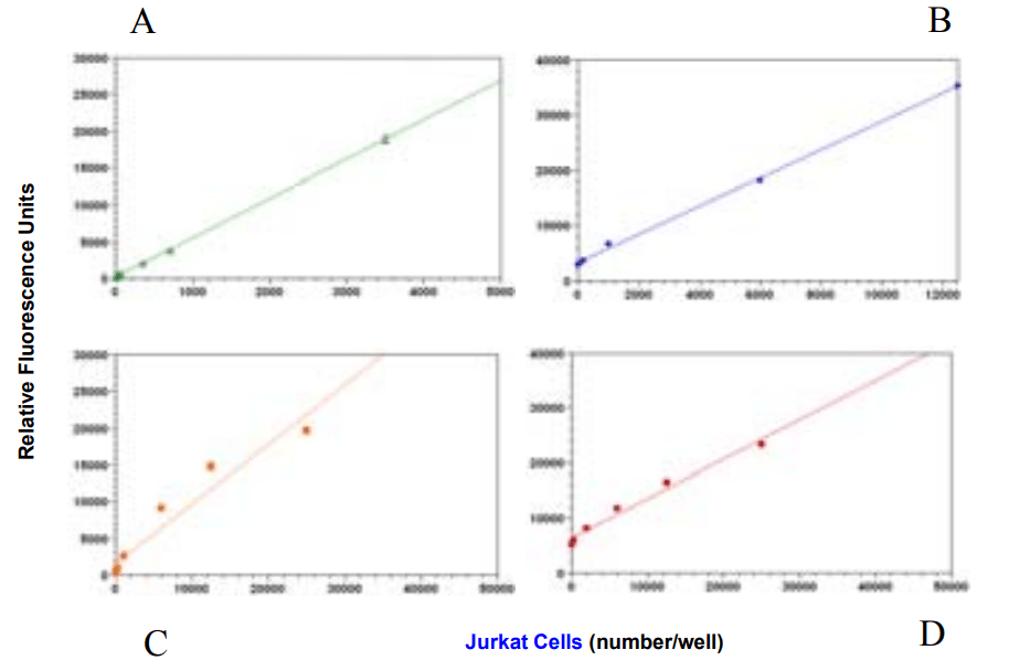
Jurkat cell number response was measured with Calcein AM, Calcein Orange™, Calcein Red™ and CytoCalcein™. Jurkat cells at 0 to 50,000 cells/well/100 µL were seeded in a 96-well black wall/clear bottom Costar plate. The cells were incubated with 5 uM each of Calcein's for 1 hr at 37oC. The fluorescence intensity was measured at Ex/Em = 490/525 nm (A, Calcein, AM), 485/540 nm (B, Calcein Orange™ ), 590/670 nm (C, Calcein Red™ ), and 405/450 nm (D, CytoCalcein™ Violet 450 ), using NOVOstar instrument (from BMG Labtech).

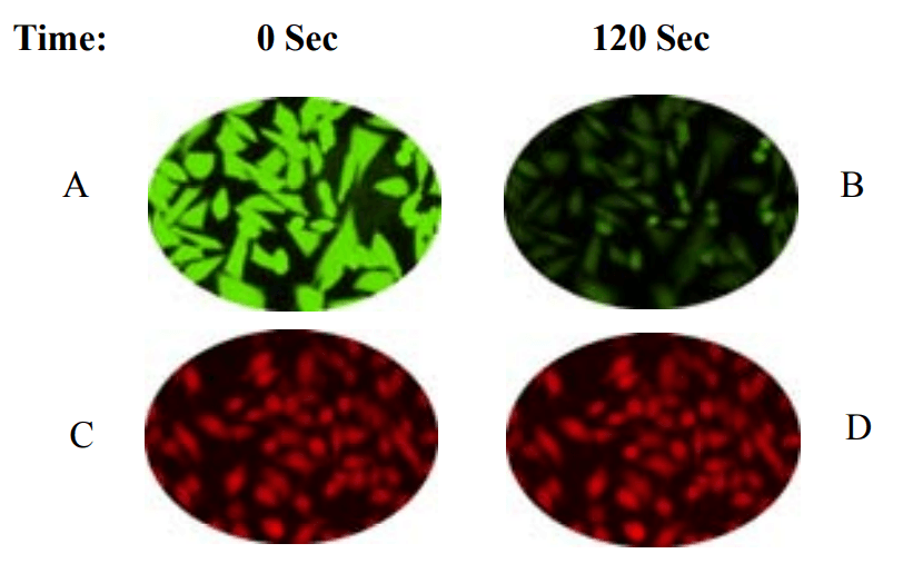
Photobleaching comparison of Calcein, AM and Calcein Red™ on CPA cells. CPA cells were plating at 30,000 cells/well in a 96-well Costar black wall/clear bottom plate for overnight, 2-5 µM of Calcein, AM or Calcein Red™ were incubated in a 37oC, 5% CO2 incubator for 30 min. The intensity of fluorescence vs time was measured with a standard fluorescence microscope (Olympus IX71). A and B: Calcein, AM at time 0 and 120 seconds exposure time (FITC filter) C and D: Calcein Red™ at time 0 and 120 seconds exposure time (Cy5 filter).

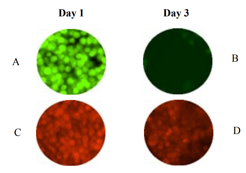
Comparison of the effect of cytotoxcity of Calcein, AM and Calcein Orange™ on Jurkat cells. Jurkat cells were dye-loaded with 5 µM of Calcein, AM or Calcein Orange™ in a 37oC, 5% CO2 incubator for 30 min, then washed with growth media. The dye-loaded cells were incubated in a 37oC, 5% CO2 incubator for 3 days. The intensity of fluorescence was measured with a standard fluorescence microscope (Olympus IX71) every day after the dye-loading. A and B: Calcein, AM at day 1 and day 3 after dye-loading (FITC filter) C and D: Calcein Orange™ at day 1 and day 3 after dye-loading (TRITC filter).

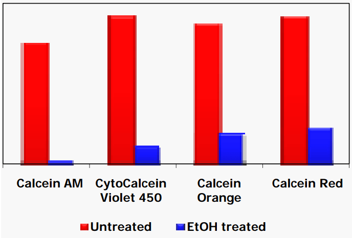
Comparison of CPA cell cytotoxicity monitored by Calcein AM, CytoCalcein™ Violet 450, Calcein Orange™ and Calcein Red™. CPA cells were treated with 25% Ethanol for 30 mins and then dye loaded with different dyes using the untreated as controls. Fluorescence were read with NOVOstar microplate reader (BMG Labtech).
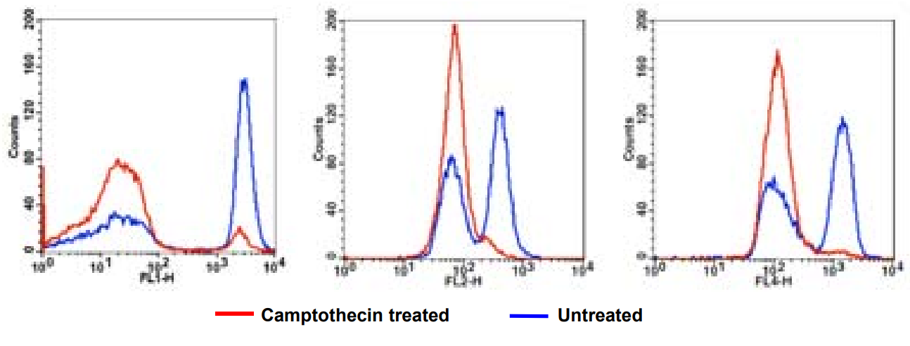
Flow Cytometric analysis on effect of camptothecin-induced Jurkat cell cytotoxity monitored by A: Calcein, AM, B: Calcein Orange™ and C: Calcein Red ™. Jurkat cells were dye-loaded for 20-30 minutes and measured with a FACSCalibur (Becton Dickinson, San Jose, CA) flow cytometer using FL1 channel ( Calcein , AM), FL2 Channel (Calcein Orange™) and FL4 channel (Calcein Red™).
Summary
CytoCalcein™, Calcein Orange™ and Calcein Red™ are novel fluorescent probes that can be used with Calcein AM or other green fluorescent dyes for multicolor analysis. They have the following benefits and features:
- Multiwavelength Analysis: CytoCalcein™ Violet 450: Ex/Em = 405/450nm Calcein Orange™: Ex/Em = 525/550nm Calcein Red™: Ex/Em = 649/650nm
- User-Friendly: CytoCalcein™ Violet 450, Calcein Orange™ and Calcein Red™ are compatible with existing settings and filter sets of fluorescence microplate readers, cell imagers and flow cytometers.
- Minimal Cytotoxitciy: Calcein Orange™ has minimal cytotoxicity even after 2 weeks dye-loading.
Original created on January 16, 2010, last updated on January 16, 2010
Tagged under: