logo
AAT Bioquest

Calcein Red™ AM

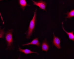
Images of Live HeLa cells stained with Calcein Red <sup>TM</sup>, AM (Cat.21900). Cell nuclei were stained with Hoechst 33342 (Blue, Cat#17535).
Images of HeLa cells stained with Calcein Red&trade; AM. Left: Live HeLa cells; Right: Fixed HeLa cells.
Ordering information
Price
Catalog Number
Unit Size
Quantity
Add to cart
Additional ordering information
Telephone1-800-990-8053
Fax1-800-609-2943
Emailsales@aatbio.com
InternationalSee distributors
Bulk requestInquire
Custom sizeInquire
ShippingStandard overnight for United States, inquire for international
Request quotation
Physical properties
Molecular weight1015.79
SolventDMSO
Spectral properties
Excitation (nm)562
Emission (nm)576
Storage, safety and handling
H-phraseH303, H313, H333
Hazard symbolXN
Intended useResearch Use Only (RUO)
R-phraseR20, R21, R22
StorageFreeze (< -15 °C); Minimize light exposure
UNSPSC12352200

OverviewpdfSDSpdfProtocol


Molecular weight
1015.79
Excitation (nm)
562
Emission (nm)
576
Calcein AM is one of the most popular fluorescent probes used for labeling and monitoring cellular functions of live cells. However, the single color of Calcein AM makes it impossible to use this valuable reagent in the multicolor applications. For example, it is impossible to use Calcein AM in combination of GFP-tranfacted cells due to the same color to GFP. To address this color limitation of Calcein AM, we have developed Calcein Orange™, Calcein Red™ and Calcein Deep Red™. These new Calcein AM analogs enable the multicolor labeling and functional analysis of live cells in combination with Calcein AM. Non-fluorescent Calcein Red™ AM can easily get into live cells and hydrolyzes to generate strongly fluorescent Calcein Red™ dye. Calcein Red™ dye can be monitored with the common TRITC/Cy3 filter set. AAT Bioquest offers Calcein Red™ as a reference dye to Calcein Red™ AM.
Platform


Flow cytometer

Excitation532/561 nm laser
Emission585/40 nm filter

Fluorescence microscope

ExcitationTRITC filter set
EmissionTRITC filter set
Recommended plateBlack wall/clear bottom

Fluorescence microplate reader

Excitation540
Emission590
Cutoff570
Recommended plateBlack wall/clear bottom
Instrument specification(s)Bottom read mode

Example protocol


PREPARATION OF STOCK SOLUTIONS

Unless otherwise noted, all unused stock solutions should be divided into single-use aliquots and stored at -20 °C after preparation. Avoid repeated freeze-thaw cycles.

Calcein Red™ AM Stock Solution
Prepare a 2 to 5 mM stock solution of Calcein Red™ AM in high-quality, anhydrous DMSO.
Note     The nonionic detergent Pluronic® F-127 can be used to increase the aqueous solubility of AM esters. In the staining buffer, the final Pluronic® F-127 concentration should be approximately 0.02%. A variety of Pluronic® F-127 products can be purchased from AAT Bioquest. Avoid long-term storage of AM esters in the presence of Pluronic® F-127.

PREPARATION OF WORKING SOLUTION

Calcein Red™ AM Working Solution
Prepare a Calcein Red™ AM working solution of 1 to 10 µM in the buffer of your choice (e.g., Hanks and Hepes buffer). For most cell lines, Calcein Red™ AM at the final concentration of 4 to 5 µM is recommended. The exact concentration of indicators required for cell loading must be determined empirically.
Note     If your cells contain organic anion-transporters, probenecid (1–2.5 mM) or sulfinpyrazone (0.1–0.25 mM) may be added to the working solution to reduce leakage of the de-esterified indicators.

SAMPLE EXPERIMENTAL PROTOCOL

  1. Prepare cells for imaging.
  2. Remove the cell culture medium and wash cells once with serum-free buffer to remove any remaining media.
    Note     Serum in cell culture media may contain esterase activity, which can increase background interference.
  3. Add Calcein Red™ AM working solution to the culture.
  4. Incubate cells at 37 °C for 30 to 60 minutes.
  5. Replace the dye working solution with HHBS or buffer of your choice (containing an anion transporter inhibitor, such as 1 mM probenecid, if applicable) to remove any excess probes.
  6. Measure the fluorescence intensity using either a fluorescence microscope equipped with a TRITC filter set, a flow cytometer equipped with green/yellow laser and a 585/40 nm filter, or a fluorescence plate reader at Ex/Em = 540/590 nm cutoff 570 nm. 

Calculators


Common stock solution preparation

Table 1. Volume of DMSO needed to reconstitute specific mass of Calcein Red™ AM to given concentration. Note that volume is only for preparing stock solution. Refer to sample experimental protocol for appropriate experimental/physiological buffers.

0.1 mg0.5 mg1 mg5 mg10 mg
1 mM98.446 µL492.228 µL984.455 µL4.922 mL9.845 mL
5 mM19.689 µL98.446 µL196.891 µL984.455 µL1.969 mL
10 mM9.845 µL49.223 µL98.446 µL492.228 µL984.455 µL

Molarity calculator

Enter any two values (mass, volume, concentration) to calculate the third.

Mass (Calculate)Molecular weightVolume (Calculate)Concentration (Calculate)Moles
/=x=

Spectrum


Open in Advanced Spectrum Viewer
spectrum

Spectral properties

Excitation (nm)562
Emission (nm)576

Images


Citations


View all 30 citations: Citation Explorer
High-Throughput Neurite Outgrowth Assay Using GFP-Labeled iPSC-Derived Neurons
Authors: Zhang, Li and Li, Shuaizhang and Xia, Menghang
Journal: Current Protocols (2022): e542
AMPA receptor and RNA processing gene dysregulation are early determinants of selective motor neuron vulnerability in a mouse model of amyotrophic lateral sclerosis
Authors: Zanganeh, Pardis F and Barton, Samantha K and Lim, Katherine and Qian, Elizabeth L and Crombie, Duncan E and Bye, Christopher R and Turner, Bradley J
Journal: Brain Communications (2022)
Copper-mediated polyurethane materials with enzyme-like catalysis for biocompatibility improvement in blood environments
Authors: Dou, Jiaxin and Li, Peichuang and Zhao, Yuancong and Zhou, Lei and Li, Xin and Wang, Jin and Huang, Nan
Journal: Biosurface and Biotribology (2021)
An automated real-time microfluidic platform to probe single NK cell heterogeneity and cytotoxicity on-chip
Authors: Subedi, Nikita and Van Eyndhoven, Laura C and Hokke, Ayla M and Houben, Lars and Van Turnhout, Mark C and Bouten, Carlijn VC and Eyer, Klaus and Tel, Jurjen
Journal: Scientific Reports (2021): 1--11
High-throughput and dosage-controlled intracellular delivery of large cargos by an acoustic-electric micro-vortices platform
Authors: Aghaamoo, Mohammad and Chen, Yu-Hsi and Li, Xuan and Garg, Neha and Jiang, Ruoyu and Lee, Abraham Phillip
Journal: bioRxiv (2021)
Inflammatory smooth muscle cells induce endothelial cell alterations to influence cerebral aneurysm progression via regulation of integrin and VEGF expression
Authors: Liu, Peixi and Shi, Yuan and Fan, Zhiyuan and Zhou, Yingjie and Song, Yaying and Liu, Yingjun and Yu, Guo and An, Qingzhu and Zhu, Wei
Journal: Cell transplantation (2019): 713--722
A perfused human blood--brain barrier on-a-chip for high-throughput assessment of barrier function and antibody transport
Authors: Wevers, Nienke R and Kasi, Dhanesh G and Gray, Taylor and Wilschut, Karlijn J and Smith, Benjamin and van Vught, Remko and Shimizu, Fumitaka and Sano, Yasuteru and K, undefined and a, Takashi and Marsh, Graham and others, undefined
Journal: Fluids and Barriers of the CNS (2018): 23
Functional imaging of neuronal activity of auditory cortex by using Cal-520 in anesthetized and awake mice
Authors: Li, Jingcheng and Zhang, Jianxiong and Wang, Meng and Pan, Junxia and Chen, Xiaowei and Liao, Xiang
Journal: Biomedical Optics Express (2017): 2599--2610
Etablierung eines 3D in vitro Blutgef{\"a}{\ss}-/Gewebemodells zur Testung spezifischer Therapeutika zur Leuk{\"a}miebehandlung
Authors: Bersi, Heidi
Journal: (2017)

References


View all 84 references: Citation Explorer
Functional evidence that the self-renewal gene NANOG regulates esophageal squamous cancer development
Authors: Li, Deng and Xiang, Xiaocong and Yang, Fei and Xiao, Dongqin and Liu, Kang and Chen, Zhu and Zhang, Ruolan and Feng, Gang
Journal: Biochemical and Biophysical Research Communications (2017)
Localized functional chemical stimulation of TE 671 cells cultured on nanoporous membrane by calcein and acetylcholine
Authors: Zibek S, Stett A, Koltay P, Hu M, Zengerle R, Nisch W, Stelzle M.
Journal: Biophys J. (2006)
A vaccination and challenge model using calcein marked fish
Authors: Klesius PH, Evans JJ, Shoemaker CA, Pasnik DJ.
Journal: Fish Shellfish Immunol (2006): 20
Novel fluorescence assay using calcein-AM for the determination of human erythrocyte viability and aging
Authors: Bratosin D, Mitrofan L, Palii C, Estaquier J, Montreuil J.
Journal: Cytometry A (2005): 78
Cytotoxic effects of 100 reference compounds on Hep G2 and HeLa cells and of 60 compounds on ECC-1 and CHO cells. I mechanistic assays on ROS, glutathione depletion and calcein uptake
Authors: Schoonen WG, Westerink WM, de Roos JA, Debiton E.
Journal: Toxicol In Vitro (2005): 505
Calcein AM release-based cytotoxic cell assay for fish leucocytes
Authors: Iwanowicz LR, Densmore CL, Ottinger CA.
Journal: Fish Shellfish Immunol (2004): 127
Calcein-AM is a detector of intracellular oxidative activity
Authors: Uggeri J, Gatti R, Belletti S, Sc and roglio R, Corradini R, Rotoli BM, Orl and ini G., undefined
Journal: Histochem Cell Biol (2004): 499
Comparison of the usefulness of the MTT, ATP, and calcein assays to predict the potency of cytotoxic agents in various human cancer cell lines
Authors: Mueller H, Kassack MU, Wiese M.
Journal: J Biomol Screen (2004): 506
In vitro assay of mineralized-tissue formation on titanium using fluorescent staining with calcein blue
Authors: Goto T, Kajiwara H, Yoshinari M, Fukuhara E, Kobayashi S, Tanaka T.
Journal: Biomaterials (2003): 3885
The effects of calcium chloride and sodium chloride on the electroporation-mediated skin permeation of fluorescein isothiocyanate (FITC)-dextrans in vitro
Authors: Tokudome Y, Sugibayashi K.
Journal: Biol Pharm Bull (2003): 1508