
Products
Services
Resources
Selection Guides
About
Endoplasmic Reticulum (ER)
The endoplasmic reticulum (ER) is an organelle present in most eukaryotic cells. It primarily synthesizes and transports cellular products such as proteins, lipids, and hormones. Structurally, the ER comprises an interconnected network of flattened, membrane-enclosed sacs and tubules extending from the nuclear membrane throughout the cytoplasm. These sac-like structures and tubules are held together and supported by the cytoskeleton.
The endoplasmic reticulum comprises two structurally and functionally distinct domains: the rough endoplasmic reticulum (rough ER) and the smooth endoplasmic reticulum (smooth ER). The rough ER is studded with ribosomes that synthesize secretory and membrane proteins. Once synthesized, membrane-bound transport vesicles shuttle these proteins to the Golgi apparatus. The smooth ER lacks ribosomes and is responsible for synthesizing lipids, phospholipids, and steroids. Secondary responsibilities of the smooth ER include carbohydrate and steroid metabolism, detoxification, and modulation of calcium ions.
ER Tracer™ dyes are membrane-permeant stains that are highly selective for the endoplasmic reticulum in living cells. As the key component of Cell Navigator® Live Cell Endoplasmic Reticulum Staining Kits, ER Tracer™ dyes can be multiplexed with other fluorescent proteins or probes in live cell multiparametric studies or after fixation for colocalization studies. ER Tracer™ stains may not bind the ER selectively in certain cell lines.
Highly selective for ER over other compartments, such as lysosomes and mitochondria | ER staining well-retained after cell fixation |
Blue, green and red fluorescence for multiplexing with other fluorescent probes | Kits include a robust staining protocol and materials for 100 tests |
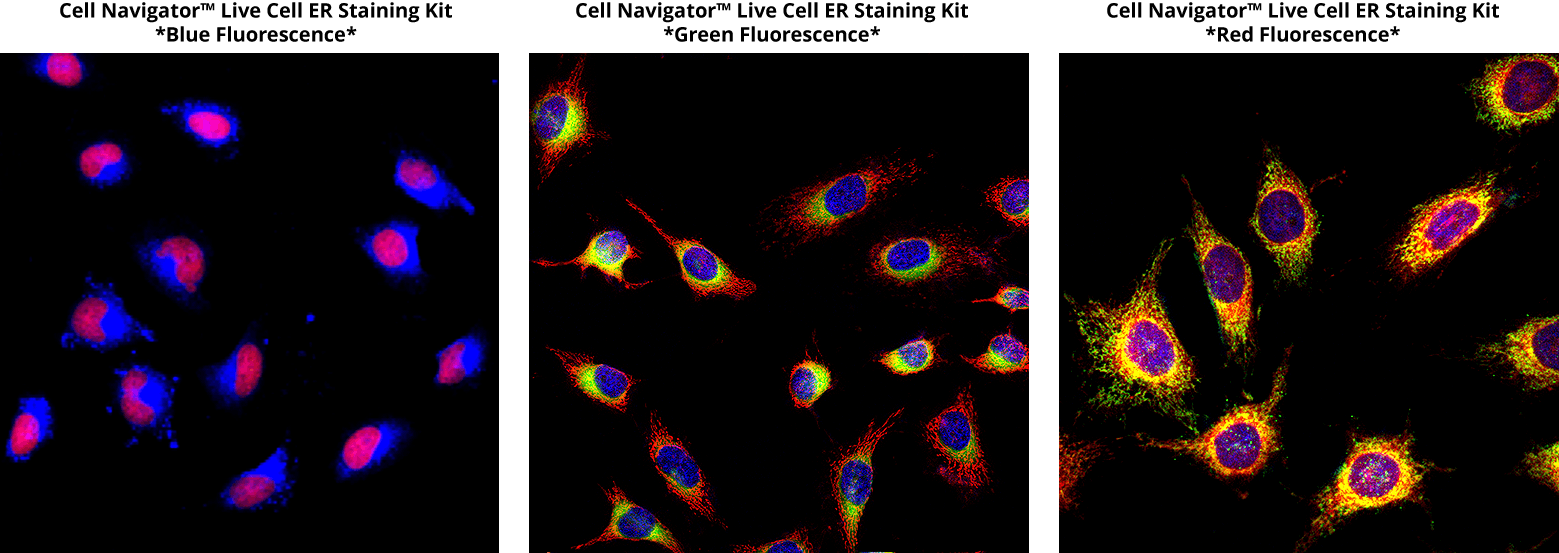 ER of HeLa cells labeled using Cell Navigator™ Live Cell Endoplasmic Reticulum Staining Kits. ER, nuclei, mitochondria, or F-actin were stained, respectively, with (Left) Cell Navigator™ Live Cell Endoplasmic Reticulum Staining Kit Blue Fluorescence (Catalog Number 22634) and Nuclear Red™ LCS1 (Catalog Number 17542), (Center) Cell Navigator™ Live Cell Endoplasmic Reticulum Staining Kit Green Fluorescence (Catalog Number 22635), DAPI (Catalog Number 17507) and Phalloidin-iFluor™ 594 (Catalog Number 23122), and (Right) Cell Navigator™ Live Cell Endoplasmic Reticulum Staining Kit Red Fluorescence (Catalog Number 22636), MitoLite™ Green (Catalog Number 22675) and Hoechst 33342 (Catalog Number 17535).
ER-Lite™ Blue-White DPX is a cell-permeable, photostable ER stain for long-term live-cell imaging of ER dynamics. It has a large Stokes shift (Ex/Em = 372/548 nm) and labels the ER with minimal off-target mitochondrial staining. ER fractions for protein analysis and glycosylation studies can be prepared with the ReadiPrep™ Endoplasmic Reticulum (ER) Isolation Kit (Catalog Number 67302).
Lipid droplets form at the ER and store neutral lipids, mainly triglycerides and cholesterol esters. Droplite™ Green, LipidLite™ Green, Bodi Fluor™ 493/503, and Nile Red stain intracellular lipid droplets for fluorescence imaging. Cell Navigator® Fluorimetric Lipid Droplet Assay Kits quantify lipid droplet accumulation with blue, green, or red fluorescence.
This document (01.0013.211015r2) was last updated on Wed Nov 11 2026. All trademarks and registered trademarks mentioned herein are the property of their respective owners.
Endoplasmic Reticulum (ER)