
Products
Services
Resources
Selection Guides
About
Nuclear and Nucleolar Stains
The cell nucleus is a membrane-bound organelle that contains most of the cell's genetic material (DNA) in the form of chromosomes. The main components of the nucleus include the nuclear envelope, a double membrane that encloses the entire organelle and isolates its contents from the cytoplasm, and the nuclear matrix, a network within the nucleus that adds mechanical support. Its primary functions include controlling gene expression, regulating DNA replication, and preserving the integrity of the DNA it houses.
The nucleus is most often visualized by fluorescence microscopy or flow cytometry with high-affinity nucleic acid dyes. Before binding to DNA or RNA, the intrinsic fluorescence of these dyes is minimal, and once bound, their fluorescence intensity increases significantly. Nucleic acid dyes are available in membrane-permeant and impermeant formats for live and dead cell analysis and quantification, and in various wavelengths to support multiplex and colocalization studies with other fluorescent probes.
| Cell permeability | Use for | |
|---|---|---|
| Live-Cell Stains | Membrane permeant | Nuclear counterstaining and morphology in living cells |
| Fixed / Dead-Cell Stains | Membrane impermeant | Dead-cell indicators and nuclear counterstains for fixed or permeabilized samples |
| Nucleolar Stains | Permeant (LCS1) or impermeant (DCS1) | Nucleolar RNA labeling with minimal nuclear and cytoplasmic background |
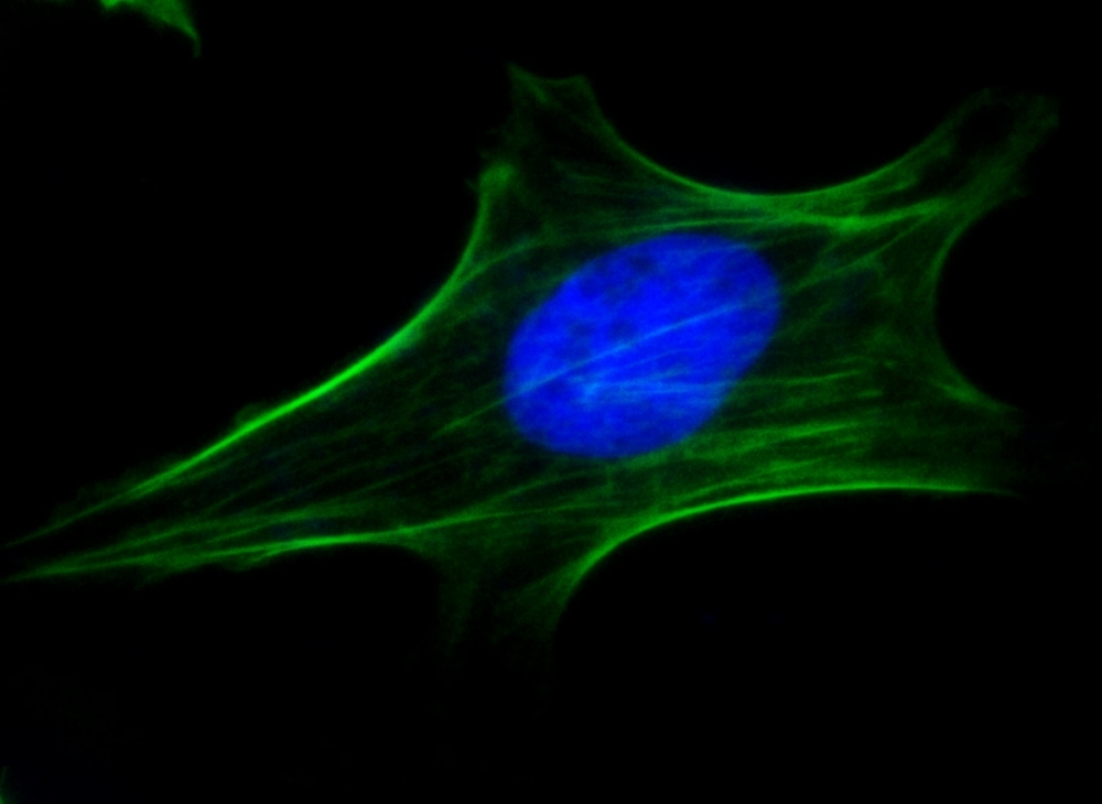
Membrane-permeant nucleic acid dyes are primarily used to label nuclei in live cells with intact plasma membranes. Not all of them stain live cells exclusively: some, like Hoechst dyes, also stain fixed or permeabilized cells. LiveONLY™ Nuclear Green (Catalog Number 17687) and LiveONLY™ Nuclear Red (Catalog Number 17688) are live-cell-only nuclear stains that do not label cells with compromised membranes.
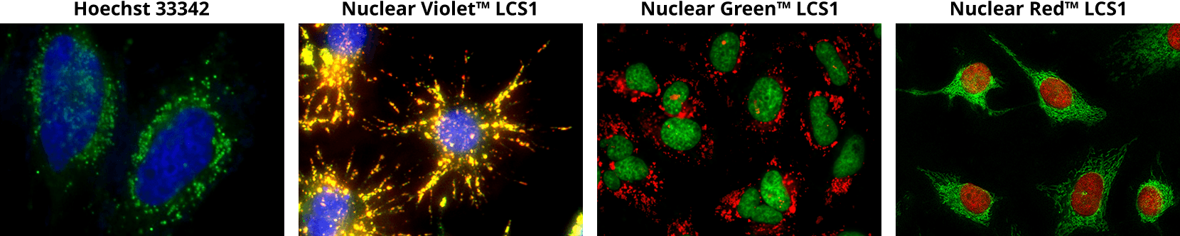
From left to right:
HeLa cell nuclei were stained with Hoechst 33342 (Catalog 17535, blue).
Live Raw 264.7 cells stained Nuclear Violet™ LCS1 (Catalog 17543, blue).
Oleic acid treated HeLa cells stained with Nuclear Green™ LCS1 (Catalog 17540, green).
Live HeLa cells stained with Nuclear Red™ LCS1 (Catalog 17542, red).
Membrane-impermeant nucleic acid dyes label the nuclei of cells with compromised plasma membranes, including dead, fixed, or permeabilized cells. Since they cannot label live cells, they are frequently used in cell viability studies as dead-cell indicators or as nuclear counterstains in fixed or permeabilized samples. Examples include DAPI, propidium iodide (PI), 7-aminoactinomycin D (7-AAD), Nuclear Blue™ DCS1, Nuclear Green™ DCS1, Nuclear Orange™ DCS1, and Nuclear Red™ DCS1.
The cyanine nucleic acid stains DiTO™, DiYO™, and TWO-PRO™ display exceptional sensitivity for nucleic acids, including double-stranded DNA (dsDNA), single-stranded DNA (ssDNA), and RNA. The symmetric cyanine dimers DiTO™ and DiYO™ exhibit a 100- to 1000-fold fluorescence enhancement upon nucleic acid binding, and their molar extinction coefficients are approximately an order of magnitude greater than those of ethidium homodimers.
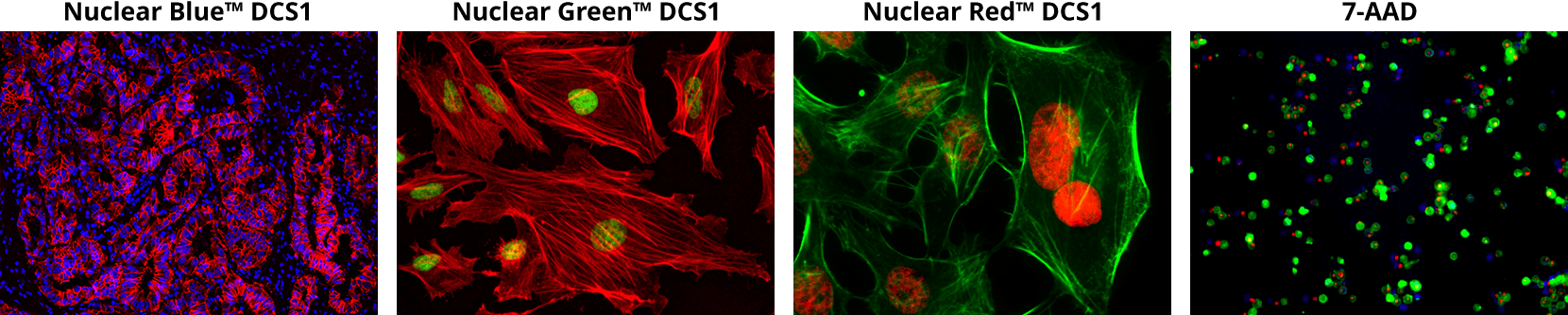
From left to right:
Paraffin-embedded human lung adenocarcinoma with nuclei stained using Nuclear Blue™ DCS1 (Catalog 17548, blue).
HeLa cells fixed with 4% formaldehyde then stained with Nuclear Green™ DCS1 (Catalog 17550, green)
HeLa cells fixed with 4% formaldehyde then stained with Nuclear Red™ DCS1 (Catalog 17552, red)
Jurkat cells stained with 7-AAD (Catalog 17501, red).
The nucleolus is a dense, membrane-less substructure within the nucleus that serves as the primary site for ribosomal RNA (rRNA) synthesis and ribosome biogenesis. Unlike the surrounding nucleoplasm, which is predominantly DNA-rich, the nucleolus is characterized by high concentrations of RNA and RNA-binding proteins. This compositional difference allows preferential visualization of the nucleolus using RNA-selective fluorescent dyes that show enhanced accumulation in RNA-rich regions.
AAT Bioquest's Nucleolus™ stains are designed to preferentially label nucleolar RNA with minimal background staining in the surrounding nuclear and cytoplasmic compartments. These dyes are particularly useful for:
- Ribosome biogenesis studies: Monitoring nucleolar morphology and activity as indicators of cellular protein synthesis capacity
- Cell stress and disease research: Nucleolar size and organization are sensitive markers of cellular stress, with alterations observed in cancer, viral infection, and neurodegenerative conditions
- Cell cycle analysis: Nucleolar dynamics change throughout the cell cycle, with disassembly during mitosis and reassembly in early G1
- Multiplexed imaging: Available in blue, green, and red for co-localization studies with DNA stains, immunofluorescence markers, or other cellular probes
This document (01.0087.211015r2) was last updated on Wed Nov 11 2026. All trademarks and registered trademarks mentioned herein are the property of their respective owners.
Nuclear and Nucleolar Stains