
Products
Services
Resources
Selection Guides
About
Plasma Membrane Stains
The plasma membrane is a thin semi-permeable membrane that separates the interior of all cells from their extracellular environment. It consists of a phospholipid bilayer embedded throughout with proteins, such as ion channels, carrier proteins, and receptor proteins, that carry out specific membrane functions. It provides structural support and protection for all cells, maintains ionic homeostasis, and is an integral component in cell adhesion, cell-cell communication, ion conductivity, and cell signaling pathways.
AAT Bioquest offers plasma membrane staining kits, fluorescent wheat germ agglutinin (WGA) and concanavalin A (ConA) conjugates, and lipophilic tracers such as DiI, DiO, DiD, and DiR for investigating lipid metabolism, imaging cell structures, and analyzing biophysical and cell signaling processes.
| How it labels | Sample | Colors | |
|---|---|---|---|
| Staining Kits | Cellpaint™ membrane stains in a ready-to-use Cell Navigator® kit | Live cells; staining is retained after formaldehyde fixation | Green, orange, and red |
| WGA and ConA Conjugates | Lectins that bind cell-surface glycans | Live or fixed cells | Blue to near-infrared (iFluor® and XFD dyes) |
| Lipophilic Tracers | Carbocyanine dyes that insert into the lipid bilayer | Live or fixed cells and tissues | Green to near-infrared |
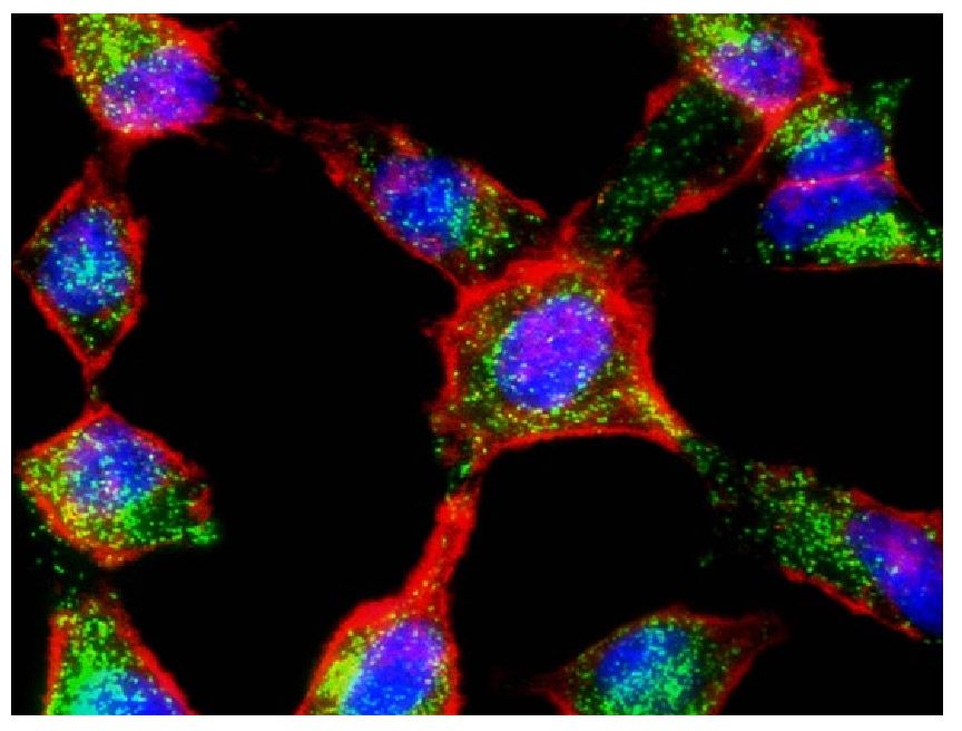
Cell Navigator® Plasma Membrane Staining Kits rapidly and uniformly label the plasma membrane in living cells without the cell-type differences exhibited by lectins. The kits employ Cellpaint™ plasma membrane stains, which are designed for live-cell labeling. The membrane staining is well preserved after fixation with formaldehyde, enabling multiplexing with other fluorescent conjugates or proteins.
Bright, photostable fluorescence with high signal-to-noise ratios | Uniform staining across a broad range of mammalian cell types, suspended or attached |
Staining well-retained in the plasma membrane after fixation | Available in green, orange and red fluorescence for multicolor staining |
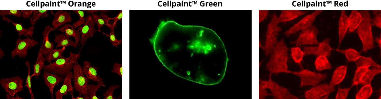 Live HeLa cells were stained with Cellpaint™ Orange, then fixed in 4% formaldehyde and costained with Nuclear Green™ DCS1. Live HL-60 cells were stained with Cellpaint™ Green. Live HeLa cells were stained with Cellpaint™ Red.
Wheat germ agglutinin (WGA) and concanavalin A (ConA) are lectins that label the plasma membrane by binding glycans on cell-surface glycoproteins and glycolipids. They recognize different sugars, so each labels a different set of surface glycans. Both stain live or fixed cells that have not been permeabilized, and both are available as iFluor® and XFD conjugates.
WGA is a ~36 kDa lectin with high affinity for N-acetyl-D-glucosamine and sialic acid residues on glycoconjugates. It is routinely used to label the cell membranes of mammalian cells, gram-positive bacteria, and yeast bud scars, as well as skeletal and cardiac sarcolemma and fibrotic scar tissue. iFluor® WGA conjugates exhibit superior brightness, photostability, and water solubility, outperforming Alexa Fluor® WGA conjugates and other spectrally similar conjugates, and are available from blue (iFluor® 350) to near-infrared (iFluor® 790).
ConA is one of the most widely used lectins in cell biology. It binds α-D-mannosyl and α-D-glucosyl residues and is used to characterize glycoproteins and other sugar-containing structures on the cell surface. iFluor® ConA conjugates are available from green (iFluor® 488) to near-infrared (iFluor® 750).
Stains non-permeabilized live or fixed cells in 10 to 30 minutes | Permeabilizing after fixation also stains Golgi and ER structures |
WGA withstands fixation and permeabilization and serves as a neuronal tracer | Range of colors for multiplex and colocalization with other conjugates or proteins |
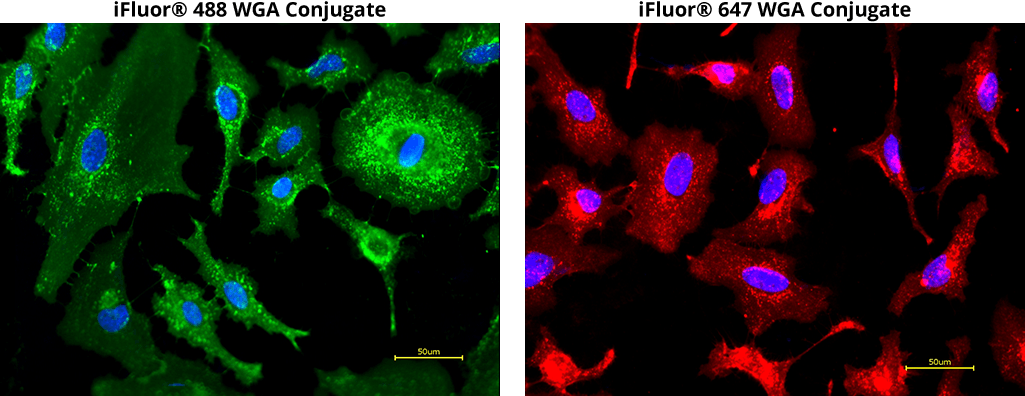 Left: Live HeLa cells were stained with iFluor® 488 WGA conjugate (green) and Hoechst 33342 (blue). Right: Live HeLa cells were stained with iFluor® 647 WGA conjugate (red) and Hoechst 33342 (blue).
DiA, DiI, DiO, DiD, and DiR are a family of dialkylcarbocyanine dyes used extensively for labeling hydrophobic structures such as cell membranes, and as anterograde or retrograde neuronal tracers in living and fixed cells and tissues. Once applied to cells, these dyes diffuse rapidly and laterally within the plasma membrane, resulting in detailed, uniform staining of the entire cell surface. Staining is specific, as these dyes do not transfer via gap junctions or to unlabeled cells unless the membrane of the labeled cell is disturbed. In addition to membrane staining and neuronal tracing, lipophilic carbocyanines have been used in cytotoxicity assays, lipoprotein labeling, cell adhesion studies, cell migration tracing, and fluorescence recovery after photobleaching (FRAP) assays.
Extremely high extinction coefficients | Polarity-dependent fluorescence |
Short excited-state lifetimes | DiO, DiI, DiD and DiR emit green, orange, red and infrared for multicolor imaging and flow |
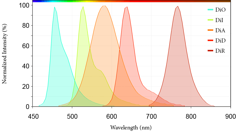 Emission spectra of representative dialkylcarbocyanine dyes spanning green to near-infrared fluorescence.
This document (01.0033.211015r2) was last updated on Wed Nov 11 2026. All trademarks and registered trademarks mentioned herein are the property of their respective owners.