logo
AAT Bioquest

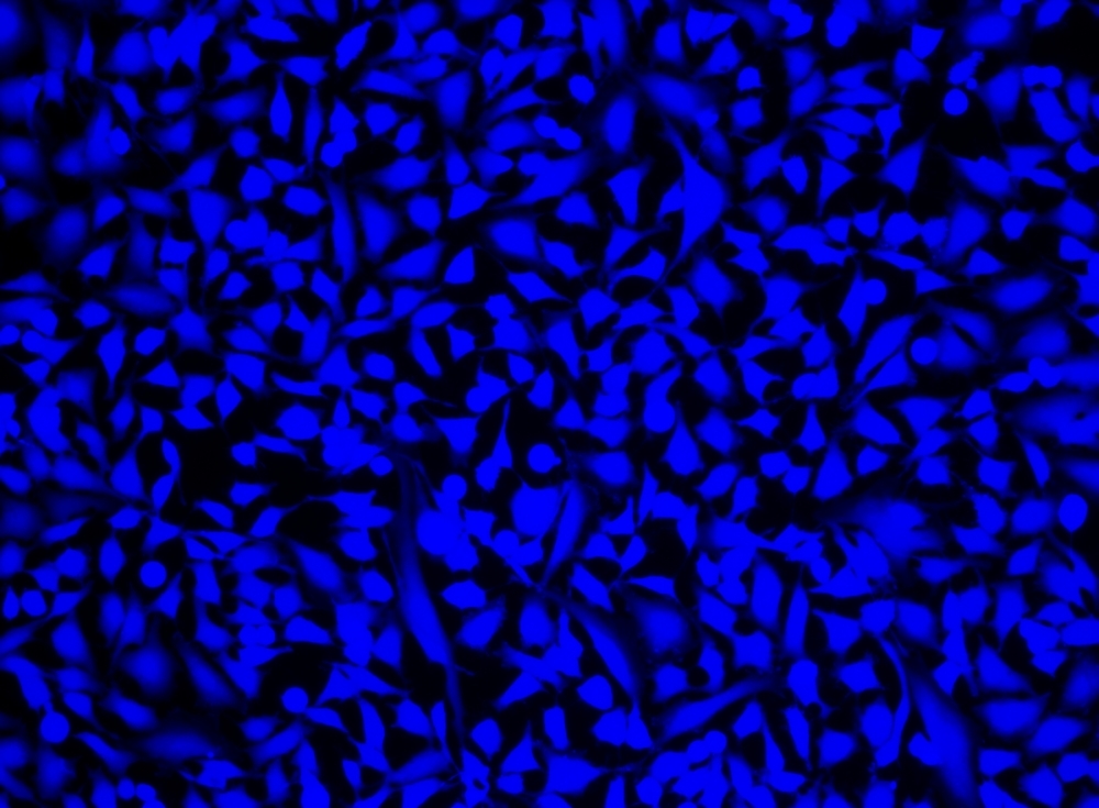
Cell Explorer™ Live Cell Labeling Kit *Blue Fluorescence*

Our Cell Explorer™ fluorescence imaging kits are a set of tools for labeling cells for fluorescence microscopic investigations of cellular functions. The effective labeling of cells provides a powerful method for studying cellular events in a spatial and temporal context. This particular kit is designed to uniformly label live cells in blue fluorescence. The kit uses a proprietary dye that gets enhanced fluorescence upon entering into live cells. The dye is a hydrophobic compound that easily permeates intact live cells. The hydrolysis of the weakly fluorescent substrate by intracellular esterases generates a strongly fluorescent hydrophilic product that is well-retained in the cell cytoplasm. Cells grown in black-walled plates can be stained and quantified in less than two hours. It can be readily adapted for high-throughput assays in a wide variety of fluorescence platforms such as microplate assays, immunocytochemistry and flow cytometry. It is useful for a variety of studies, including cell adhesion, chemotaxis, multidrug resistance, cell viability, apoptosis and cytotoxicity. The kit provides all the essential components with an optimized cell-labeling protocol.

Example protocol

AT A GLANCE

Protocol summary

  1. Prepare cells in growth medium
  2. Remove the medium
  3. Add Calcein UltraBlue™ working solution (100 µL/well for 96-well plates or 25 µL/well for 384-well plates)
  4. Incubate the cells at 37 oC for 30 minutes to 2 hours
  5. Wash the cells
  6. Examine the specimen under under fluorescence microscope with DAPI filter (Ex/Em = 360/445 nm)

Important notes
Thaw all the components at room temperature before starting the experiment.

PREPARATION OF STOCK SOLUTION

Unless otherwise noted, all unused stock solutions should be divided into single-use aliquots and stored at -20 °C after preparation. Avoid repeated freeze-thaw cycles.

1. Calcein UltraBlue™ stock solution:
Add 20 µL of DMSO into the vial of Calcein UltraBlue™ (Component A) and mix well to make Calcein UltraBlue™ stock solution. Protect from light. Note: 10 µL of Calcein UltraBlue™ stock solution is enough for 1 plate. For storage, seal tubes tightly.

PREPARATION OF WORKING SOLUTION

Add 10 µL of Calcein UltraBlue™ stock solution into 10 mL of HHBS (Component B) and mix well to make Calcein UltraBlue™ working solution. Note: Protect from light.  

For guidelines on cell sample preparation, please visit
https://www.aatbio.com/resources/guides/cell-sample-preparation.html

SAMPLE EXPERIMENTAL PROTOCOL

  1. Remove the growth medium from the cell plates.

  2. Add 100 µL/well (96-well plate) or 25 µL/well (384-well plate) of Calcein UltraBlue™ working solution into the cell plate.

  3. Incubate the cells in a 37°C, 5% CO2 incubator for 30 minutes to 2 hours.

  4. Wash the cells with HHBS (Component B), and add growth medium or HHBS back to the cells.

  5. Image the cells using a fluorescence microscope with DAPI filter (Ex/Em = 360/445 nm).

Citations

View all 6 citations: Citation Explorer
Development of a novel senolysis approach targeting the senescent fibroblast marker HTR2A via antibody-dependent cellular cytotoxicity.
Authors: Takaya, Kento and Asou, Toru and Kishi, Kazuo
Journal: Rejuvenation Research (2023)
The preparation of hollow mesoporous bioglass nanoparticles with excellent drug delivery capacity for bone tissue regeneration
Authors: Wang, Yudong and Pan, Haobo and Chen, Xiaofeng
Journal: Frontiers in chemistry (2019): 283
Autophagy proteins are not universally required for phagosome maturation
Authors: Cemma, Marija and Grinstein, Sergio and Brumell, John H
Journal: Autophagy (2016): 1440--1446
Size control and biological properties of monodispersed mesoporous bioactive glass sub-micron spheres
Authors: Hu, Qing and Li, Yuli and Miao, Guohou and Zhao, Naru and Chen, Xiaofeng
Journal: Rsc Advances (2014): 22678--22687
Differential detection of tumor cells using a combination of cell rolling, multivalent binding, and multiple antibodies
Authors: Myung, Ja Hye and Gajjar, Khyati A and Chen, Jihua and Molokie, Robert E and Hong, Seungpyo
Journal: Analytical chemistry (2014): 6088--6094

References

View all 26 references: Citation Explorer
Requirements, features, and performance of high content screening platforms
Authors: Gough AH, Johnston PA.
Journal: Methods Mol Biol (2007): 41
A pharmaceutical company user's perspective on the potential of high content screening in drug discovery
Authors: Hoffman AF, Garippa RJ.
Journal: Methods Mol Biol (2007): 19
Optimizing the integration of immunoreagents and fluorescent probes for multiplexed high content screening assays
Authors: Giuliano KA., undefined
Journal: Methods Mol Biol (2007): 189
Past, present, and future of high content screening and the field of cellomics
Authors: Taylor DL., undefined
Journal: Methods Mol Biol (2007): 3
High concordance of drug-induced human hepatotoxicity with in vitro cytotoxicity measured in a novel cell-based model using high content screening
Authors: O'Brien P J, Irwin W, Diaz D, Howard-Cofield E, Krejsa CM, Slaughter MR, Gao B, Kaludercic N, Angeline A, Bernardi P, Brain P, Hougham C.
Journal: Arch Toxicol (2006): 580
Page updated on October 23, 2024

Ordering information

Price
Unit size
Catalog Number22606
Quantity
Add to cart

Additional ordering information

Telephone1-800-990-8053
Fax1-800-609-2943
Emailsales@aatbio.com
InternationalSee distributors
Bulk requestInquire
Custom sizeInquire
Technical SupportContact us
Purchase orderSend to sales@aatbio.com
ShippingStandard overnight for United States, inquire for international
Request quotation

Storage, safety and handling

H-phraseH303, H313, H333
Hazard symbolXN
Intended useResearch Use Only (RUO)
R-phraseR20, R21, R22
UNSPSC12352200

Platform

Fluorescence microscope

ExcitationDAPI filter set
EmissionDAPI filter set
Recommended plateBlack wall, clear bottom

Components