Cell Meter™ Live Cell Caspase 3/7 and Phosphatidylserine Detection Kit *Triple Fluorescence Colors*
Ordering information
Price | |
Catalog Number | |
Unit Size | |
Quantity |
Additional ordering information
Telephone | 1-800-990-8053 |
Fax | 1-800-609-2943 |
sales@aatbio.com | |
Quotation | Request |
International | See distributors |
Shipping | Standard overnight for United States, inquire for international |
Storage, safety and handling
H-phrase | H303, H313, H333 |
Hazard symbol | XN |
Intended use | Research Use Only (RUO) |
R-phrase | R20, R21, R22 |
UNSPSC | 12352200 |
Related products
Overview | ![]() ![]() |
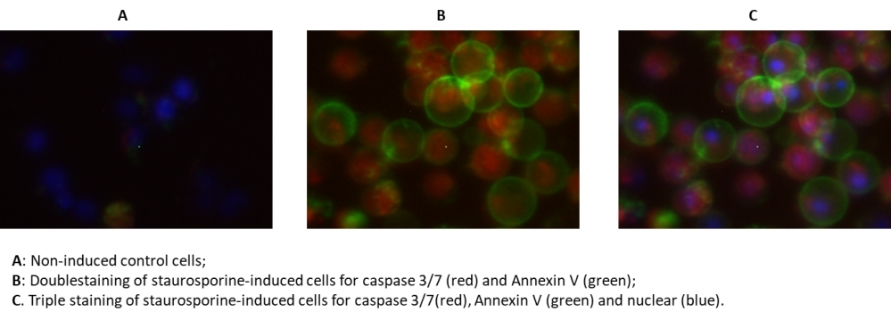
Our Cell Meter™ assay kits are a set of tools for monitoring cellular functions. In the process of apoptosis, one of key events is the activation of caspases. The activation of caspase 3/7 is an important for the initiation of apoptosis. It has been proven that caspase 3/7 has substrate selectivity for the peptide sequence Asp-Glu-Val-Asp (DEVD). This kit uses SR-DEVD-FMK as a fluorescent indicator to detect caspase 3/7 activities. SR-DEVD-FMK is cell permeable and nontoxic, once bound to caspases, the fluorescent reagent is retained inside the cell. The binding event prevents the caspases from further catalysis but will not stop apoptosis from proceeding. SR-DEVD-FMK is a red label reagent. Annexins are a family of proteins that bind to phospholipid membranes in the presence of calcium. Annexin V is used to detect apoptotic cells that express phosphatidylserine (PS) on the cell surface. The appearance of PS on the cell surface is a universal indicator of the initial/intermediate stages of cell apoptosis. Annexin V-dye conjugates monitor cell apoptosis through measuring the translocation of PS. The Annexin V-iFluor 488™ used in this kit is a green labeling reagent, with Ex/Em = 490/525 nm. The kit is designed to detect apoptosis by simultaneously monitoring Caspase 3/7 and Annexin V activities in mammalian cells. The kit also provides a Hoechst dye for labeling the nucleus of the whole population of the cells, and propidium iodide dye for staining necrosis cells. This kit is applicable for fluorescence microscope, flow cytometer, and fluorescence microplate reader. The kit provides all the essential components with an optimized assay protocol.
Platform
Flow cytometer
Excitation | 488 nm laser |
Emission | 530/30 nm, 575/26 nm, 610/20 nm filter |
Instrument specification(s) | FITC, PE, PE-Texas Red channel |
Fluorescence microscope
Recommended plate | Black wall/clear bottom |
Instrument specification(s) | FITC channel for Annexin V-iFluor 488™, TRITC channel for TF3-DEVD-FMK, |
Fluorescence microplate reader
Excitation | 490 nm, 550 nm |
Emission | 525 nm, 595 nm |
Cutoff | 515 nm, 570 nm |
Recommended plate | Black wall/clear bottom |
Instrument specification(s) | Bottom read mode |
Components
Example protocol
AT A GLANCE
Protocol summary
- Prepare cells with test compounds at a density of 2 × 10⁶ cells/mL
- Add TF3-DEVD-FMK at 1:150 ratio and/or Annexin V-iFluor 488™ into cell solution at 1:100 ratio
- Incubate the cells in a 37°C, 5% CO2 incubator for 1 hour.
- Pellet the cells, wash and resuspend the cells with buffer or growth medium
- Monitor fluorescence intensity (bottom read mode) at Ex/Em = 490/525 nm (Cutoff = 515 nm) and/or 550/595 nm (Cutoff = 570 nm), fluorescence microscope with FITC and TRITC filters, or flow cytometer with FL1 and FL2 channels for Annexin V-iFluor 488™ and TF3-DEVD-FMK respectively
Important notes
Thaw all the components at room temperature before starting the experiment.
PREPARATION OF STOCK SOLUTION
Unless otherwise noted, all unused stock solutions should be divided into single-use aliquots and stored at -20 °C after preparation. Avoid repeated freeze-thaw cycles.
1. TF3-DEVD-FMK stock solution (150X):
Add 200 µL of DMSO into the vial of TF3-DEVD-FMK (Component A) to make 150X TF3-DEVD-FMK stock solution.
For guidelines on cell sample preparation, please visit
https://www.aatbio.com/resources/guides/cell-sample-preparation.html
SAMPLE EXPERIMENTAL PROTOCOL
- Culture cells to a density optimal for apoptosis induction according to your specific induction protocol, but not to exceed 2 x 10⁶ cells/ mL (or not to exceed 3 x 10⁵ cells/100 µL/well in a 96-well black clear-bottom plate). At the same time, culture a non-induced negative control cell population at the same density as the induced population for every labeling condition. Here are a few examples for inducing apoptosis in suspension culture:
- Treating Jurkat cells with 2 µg/ml camptothecin for 3 hours.
- Treating Jurkat cells with 1 µM staurosporine for 3 hours.
- Treating HL-60 cells with 4 µg/ml camptothecin for 4 hours.
- Treating HL-60 cells with 1 µM staurosporine for 4 hours. Note: Each cell line should be evaluated on an individual basis to determine the optimal cell density for apoptosis induction.
- Treating Jurkat cells with 2 µg/ml camptothecin for 3 hours.
- Add 150X TF3-DEVD-FMK stock solution at a 1:150 ratio and/or Annexin V-iFluor 488™ (Component B) at 1:100 ratio into each well.
- Incubate the cells in a 37°C, 5% CO2 incubator for 1 hour. Note: The cells can be concentrated up to ~ 5 X 10⁶ cells/mL for TF3-DEVD-FMK labeling. For adherent cells, gently lift the cells with 0.5 mM EDTA to keep the cells intact, and wash the cells once with serum-containing media prior to incubation with TF3 -DEVD-FMK. The appropriate incubation time depends on the individual cell type and cell concentration used. Optimize the incubation time for each experiment. Annexin V flow cytometric analysis on adherent cells is not routinely tested since specific membrane damage may occur during cell detachment or harvesting. However, methods for utilizing Annexin V for flow cytometry on adherent cell types have been previously reported by Casiola-Rosen et al. and van Engelend et al.
- If desired, label the cells with a DNA stain (such as Hoechst for whole population of the cell nucleus stain, or propidium iodide for dead cells if the cells label with Annexin V-iFluor 488™ only).
- Spin down the cells at ~200g for 2 minutes and wash cells with 1 mL (or 200 µL/well if using 96-well plate) Washing Buffer (Component E) twice. Resuspend the cells in desired amount of washing buffer. Note: TF3-DEVD-FMK and Annexin V-iFluor 488™ are fluorescent, thus it is important to wash out any unbound reagent to eliminate the background. For detached cells, the concentration of cells should be adjusted to 2-5 X 10⁵ cells/100 µL aliquot per microtiter plate well.
- Monitor the fluorescence intensity by fluorescence microscope, flow cytometer, or fluorescence microplate reader at Ex/Em = 550/595 nm for TF3-DEVD-FMK, 490/525 for Annexin V-iFluor 488™, 350/461 nm for Hoechst stain, and 535/635 for propidium iodide.
For flow cytometry: Monitor the fluorescence intensity using FL1 channel for Annexin V-iFluor 488™, FL2 channel for TF3-DEVD-FMK. Gate on the cells of interest, excluding debris.
For fluorescence microscope: Place 100 µL of the cell suspensions into each of wells of a 96-well black microtiter plate. Observe cells under a fluorescence microscope using TRITC channel for TF3-DEVD-FMK, and/or FITC channel for Annexin V-iFluor 488™ (TRITC channel for propidium iodide staining, DAPI channel for Hoechst staining).
For fluorescence microplate reader: Place 100 µL of the cell suspensions into each of wells of a 96-well black microtiter plate. Monitor the fluorescence intensity (bottom read mode) with a fluorescence microplate reader at Ex/Em = 490/525 nm (Cutoff = 515 nm) for Annexin V-iFluor 488™, and/or 550/595 nm (Cutoff = 570 nn) for TF3-DEVD-FMK. Note: If it is necessary to equilibrate the cell concentrations, adjust the suspension volume for the induced cells to approximate the cell density of the non-induced population. This adjustment step is optional if your cell treatment does not result in a dramatic loss in stimulated cell population numbers.
Images

Figure 1. The fluorescence image analysis indicated the increased expression of caspase 3/7 (red, stained by TF3- DEVD-FMK) and Annexin V (Green, stained by Annexin V-iFluor 488™) in Jurkat cells induced by 1 μM staurosporine for 3 hour. The fluorescence images of the cells (300,000 cells/ well) were taken with Olympus fluorescence microscope through the DAPI, FITC, and TRITC channel respectively. Individual images taken from each channel from the same cell population were merged as shown above. A: Non-induced control cells; B: Doublestaining of staurosporine-induced cells for caspase 3/7 (red) and Annexin V (green); C: Triple staining of staurosporine-induced cells for caspase 3/7(red), Annexin V (green) and nuclear (blue).
Citations
View all 6 citations: Citation Explorer
The Effect of Therapeutic Klotho Protein on Cell Viability in HT-29 Cell Line
Authors: Sariboyaci, Ayla Eker and Uysal, Onur and Soykan, Merve Nur and Gunes, Sibel
Journal: (2022)
Authors: Sariboyaci, Ayla Eker and Uysal, Onur and Soykan, Merve Nur and Gunes, Sibel
Journal: (2022)
Enhancement of Apo2L/TRAIL signaling pathway receptors by the activation of Klotho gene with CRISPR/Cas9 in Caco-2 colon cancer cells
Authors: Gunes, Sibel and Soykan, Merve Nur and Sariboyaci, Ayla Eker and Uysal, Onur and Sevimli, Tugba Semerci
Journal: Medical Oncology (2021): 1--10
Authors: Gunes, Sibel and Soykan, Merve Nur and Sariboyaci, Ayla Eker and Uysal, Onur and Sevimli, Tugba Semerci
Journal: Medical Oncology (2021): 1--10
Helicobacter pylori secreted protein HP1286 triggers apoptosis in macrophages via TNF-independent and ERK MAPK-dependent pathways
Authors: Tavares, Raquel and Pathak, Sushil Kumar
Journal: Frontiers in Cellular and Infection Microbiology (2017): 58
Authors: Tavares, Raquel and Pathak, Sushil Kumar
Journal: Frontiers in Cellular and Infection Microbiology (2017): 58
Anthocyanin-rich blackcurrant extract inhibits proliferation of the MCF10A healthy human breast epithelial cell line through induction of G0/G1 arrest and apoptosis
Authors: Nanashima, Naoki and Horie, Kayo and Chiba, Mitsuru and Nakano, Manabu and Maeda, Hayato and Nakamura, Toshiya
Journal: Molecular Medicine Reports (2017): 6134--6141
Authors: Nanashima, Naoki and Horie, Kayo and Chiba, Mitsuru and Nakano, Manabu and Maeda, Hayato and Nakamura, Toshiya
Journal: Molecular Medicine Reports (2017): 6134--6141
Clusterin signals via ApoER2/VLDLR and induces meiosis of male germ cells
Authors: Riaz, Muhammad Assad and Stammler, Angelika and Borgers, Mareike and Konrad, Lutz
Journal: American Journal of Translational Research (2017): 1266
Authors: Riaz, Muhammad Assad and Stammler, Angelika and Borgers, Mareike and Konrad, Lutz
Journal: American Journal of Translational Research (2017): 1266
Detecting Apoptosis, Autophagy, and Necrosis
Authors: Coleman, Jack and Liu, Rui and Wang, Kathy and Kumar, Arun
Journal: Apoptosis Methods in Toxicology (2016): 77--92
Authors: Coleman, Jack and Liu, Rui and Wang, Kathy and Kumar, Arun
Journal: Apoptosis Methods in Toxicology (2016): 77--92
References
View all 71 references: Citation Explorer
Synergistic effects and mechanisms of combined tumor necrosis factor-related apoptosis-inducing ligand and chemotherapeutic drugs or radiotherapy in killing laryngeal squamous carcinoma cells in vitro
Authors: Zhang M, Zhou L.
Journal: Zhonghua Er Bi Yan Hou Tou Jing Wai Ke Za Zhi (2009): 565
Authors: Zhang M, Zhou L.
Journal: Zhonghua Er Bi Yan Hou Tou Jing Wai Ke Za Zhi (2009): 565
Down-regulation of myeloid cell leukemia 1 by epigallocatechin-3-gallate sensitizes rheumatoid arthritis synovial fibroblasts to tumor necrosis factor alpha-induced apoptosis
Authors: Ahmed S, Silverman MD, Marotte H, Kwan K, Matuszczak N, Koch AE.
Journal: Arthritis Rheum (2009): 1282
Authors: Ahmed S, Silverman MD, Marotte H, Kwan K, Matuszczak N, Koch AE.
Journal: Arthritis Rheum (2009): 1282
Agmatine protects cultured retinal ganglion cells from tumor necrosis factor-alpha-induced apoptosis
Authors: Hong S, Kim CY, Lee JE, Seong GJ.
Journal: Life Sci (2009): 28
Authors: Hong S, Kim CY, Lee JE, Seong GJ.
Journal: Life Sci (2009): 28
Exaggerated up-regulation of tumor necrosis factor alpha-dependent apoptosis in the older mouse liver following reperfusion injury: targeting liver protective strategies to patient age
Authors: Selzner M, Selzner N, Chen L, Borozan I, Sun J, Xue-Zhong M, Zhang J, McGilvray ID.
Journal: Liver Transpl (2009): 1594
Authors: Selzner M, Selzner N, Chen L, Borozan I, Sun J, Xue-Zhong M, Zhang J, McGilvray ID.
Journal: Liver Transpl (2009): 1594
Oxidant stress-induced liver injury in vivo: role of apoptosis, oncotic necrosis, and c-Jun NH2-terminal kinase activation
Authors: Hong JY, Lebofsky M, Farhood A, Jaeschke H.
Journal: Am J Physiol Gastrointest Liver Physiol (2009): G572
Authors: Hong JY, Lebofsky M, Farhood A, Jaeschke H.
Journal: Am J Physiol Gastrointest Liver Physiol (2009): G572
A pharmaceutical preparation of Salvia miltiorrhiza protects cardiac myocytes from tumor necrosis factor-induced apoptosis and reduces angiotensin II-stimulated collagen synthesis in fibroblasts
Authors: Ling S, Luo R, Dai A, Guo Z, Guo R, Komesaroff PA.
Journal: Phytomedicine (2009): 56
Authors: Ling S, Luo R, Dai A, Guo Z, Guo R, Komesaroff PA.
Journal: Phytomedicine (2009): 56
Increased apoptosis in HepG2.2.15 cells with hepatitis B virus expression by synergistic induction of interferon-gamma and tumour necrosis factor-alpha
Authors: Shi H, Guan SH.
Journal: Liver Int (2009): 349
Authors: Shi H, Guan SH.
Journal: Liver Int (2009): 349
Outside-to-inside signaling through transmembrane tumor necrosis factor reverses pathologic interleukin-1beta production and deficient apoptosis of rheumatoid arthritis monocytes
Authors: Meusch U, Rossol M, Baerwald C, Hauschildt S, Wagner U.
Journal: Arthritis Rheum (2009): 2612
Authors: Meusch U, Rossol M, Baerwald C, Hauschildt S, Wagner U.
Journal: Arthritis Rheum (2009): 2612
Tumor necrosis factor inhibitors block apoptosis of human epithelial cells of the salivary glands
Authors: Sisto M, D'Amore M, Caprio S, Mitolo V, Scagliusi P, Lisi S.
Journal: Ann N Y Acad Sci (2009): 407
Authors: Sisto M, D'Amore M, Caprio S, Mitolo V, Scagliusi P, Lisi S.
Journal: Ann N Y Acad Sci (2009): 407
Susceptibility of the C2 canine mastocytoma cell line to the effects of tumor necrosis factor-related apoptosis-inducing ligand (TRAIL)
Authors: Elders RC, Baines SJ, Catchpole B.
Journal: Vet Immunol Immunopathol (2009): 11
Authors: Elders RC, Baines SJ, Catchpole B.
Journal: Vet Immunol Immunopathol (2009): 11
Application notes
FAQ
Do you offer any fluorimetric assays that measure caspase activation/activity in live cells using a flow cytometer?
Are inflammasomes and caspase-1 related?
Does pH and staining temperature affect Annexin V-Phosphatidylserine binding?
Does propidium iodide stain apoptotic cells?
How can I tell if my cell sample is dying?
Are inflammasomes and caspase-1 related?
Does pH and staining temperature affect Annexin V-Phosphatidylserine binding?
Does propidium iodide stain apoptotic cells?
How can I tell if my cell sample is dying?
AssayWise
Intracellular pH Measurement with Dual Excitation Fluorescence Sensor BCFL
Nucleic Acid Detection, Quantification and Imaging
A practical guide for use of PE and APC in flow cytometry
Fluorescent Phalloidin: A Practical Stain for Visualizing Actin Filaments
Power Styramide™ Signal Amplification a Superior Alternative to Tyramide Signal Amplification
Nucleic Acid Detection, Quantification and Imaging
A practical guide for use of PE and APC in flow cytometry
Fluorescent Phalloidin: A Practical Stain for Visualizing Actin Filaments
Power Styramide™ Signal Amplification a Superior Alternative to Tyramide Signal Amplification