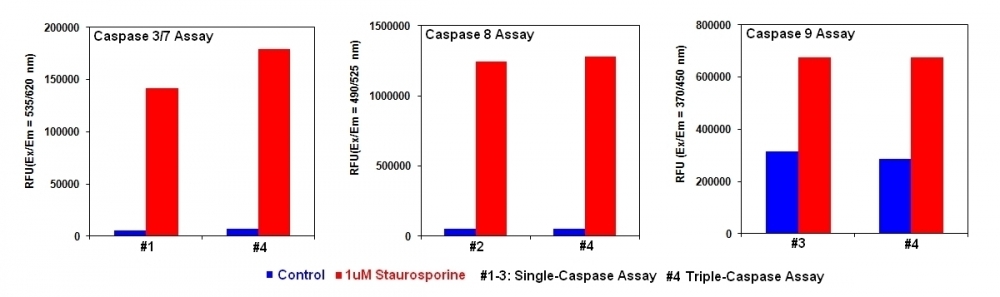
Cell Meter™ Multiplexing Caspase 3/7, 8 and 9 Activity Assay Kit *Triple Fluorescence Colors*
Overview | ![]() ![]() |
Our Cell Meter™ assay kits are a set of tools for monitoring cell viability. There are a variety of parameters that can be used for monitoring cell viability. Caspases activation is widely accepted as a reliable indicator for cell apoptosis. This particular kit is designed to simultaneously monitor four key caspases (caspase-3/7, 8 and 9) activation involved in cell apoptosis using three distinct fluorescent colors. This kit uses DEVD-ProRed™, IETD-R110 and LEHD-AMC as fluorogenic indicators for caspase 3/7, 8 and 9 activity respectively. Upon caspase cleavages, DEVD-ProRed™, IETD-R110 and LEHD-AMC caspase substrates generate three distinct fluorophores: ProRed™ (red fluorescence), R110 (green fluorescence) and AMC (blue fluorescence), which can be readily monitored in a single assay due to their nice spectral separation.
Platform
Fluorescence microplate reader
Excitation | See Table 1 |
Emission | See Table 1 |
Recommended plate | Black wall/clear bottom |
Instrument specification(s) | Use either top or bottom read mode |
Components
Component A: Caspase 3/7 Substrate (DEVD-ProRed™, 200X ) | 1 vial (50 µL/vial) |
Component B: Caspase 8 Substrate (IETD-R110, 200X) | 1 vial (50 µL/vial) |
Component C: Caspase 9 Substrate (LEHD-AMC, 200X) | 1 vial (50 µL/vial) |
Component D: Assay Buffer | 1 bottle (30 mL) |
Example protocol
AT A GLANCE
Protocol summary
- Prepare cells with test compounds
- Add equal volume of caspase working solution
- Incubate at room temperature for 30 min to 1 hour
- Monitor fluorescence intensity
Important notes
Thaw kit components at room temperature before use.
PREPARATION OF WORKING SOLUTION
1. Single caspase activity working solution
Make caspase 3/7, caspase 8 or caspase 9 working solution by adding 50 µL of substrate (Component A, B or C) into 10 mL of Assay Buffer (Component D), and mix them well.
2. Dual- or tri- caspase activity working solution:
Add 50 µL of each interested caspase substrate into 10 mL of Assay Buffer (Component D) together to make the working solution. Note: Please prepare the tested substrate solutions and the needed volume proportionally.
For guidelines on cell sample preparation, please visit
https://www.aatbio.com/resources/guides/cell-sample-preparation.html
SAMPLE EXPERIMENTAL PROTOCOL
- Treat cells by adding 10 µL/well of 10X test compounds (96-well plate) or 5 µL/well of 5X test compounds (384-plate) into PBS or the desired buffer. For blank wells (medium without the cells), add the same amount of compound buffer.
- Incubate the cell plate in a 37°C, 5% CO2, incubator for a desired period of time (3 - 5 hours for Jurkat cells treated with staurosporine) to induce apoptosis.
- Add 100 µL/well/96-well or 25 µL/well/384-well plate of desired caspase assay working solution.
- Incubate the plate at room temperature for at least 30 to 60 min, protected from light. Note: If desired, add 1 µL of 1 mM caspase inhibitor to selected samples 10 minutes before adding the assay loading solution at room temperature to confirm the inhibition of caspase activities.
- Monitor the fluorescence intensity as indicated in the table with either top or bottom read mode. Note: Sometimes, bottom read gives better signal to background ratio, centrifuge cell plate (especially for the nonadherent cells) at 800 rpm for 2 minutes (brake off) if using bottom read mode.
Table 1. Spectral wavelengths for Caspases 3/7, 8, and 9.
Capase | Fluorescence | Excitation | Emission |
Caspase 3/7 | Red | 535 nm | 620 nm |
Caspase 8 | Green | 490 nm | 525 nm |
Caspase 9 | Blue | 370 nm | 450 nm |
Images
Figure 1. Detection of Caspase Activities in Jurkat cells. Jurkat cells were seeded on the same day at 200,000 cells/well in a Costar black wall/clear bottom 96-well plate. The cells were treated with staurosporine at the final concentration of 1 mM for 4 hours (Red Bar) while the untreated cells were used as control (Blue Bar). The single-caspase assay loading solution (100 uL/well) was added (in #1 for caspase 3/7, #2 for caspase 8 or #3 for caspase 9) or Triple-caspase assay loading solution (#4 for caspase 3/7, 8 and 9 together) was added, and incubated at room temperature for 1 hour. The fluorescence intensity was measured with FlexStation fluorescence microplate reader at the indicated wavelength. The caspase 3/7, 8 and 9 activities can be detected in a single assay without interferences from other caspases.
Figure 2. cSBL induced apoptosis in H2452 and MSTO cells via activation of the caspase pathway. Caspase-3, -8, and -9 activation was detected by western blotting (A, B) or fluorometry (C, D). Fluorometry was performed independently three times and data are expressed as the mean ± SD. The statistical significance of these experiments compared with the control is shown in as follows: *P<0.05, **P<0.01, ***P<0.001. Source: Sialic acid-binding lectin from bullfrog eggs inhibits human malignant mesothelioma cell growth in vitro and in vivo by Tatsuta T et al., PLOS, Jan. 2018.
Citations
View all 23 citations: Citation Explorer
Asparagus officinalis combined with paclitaxel exhibited synergistic anti-tumor activity in paclitaxel-sensitive and-resistant ovarian cancer cells
Authors: Zhang, Xin and Wang, Jiandong and Fan, Yali and Zhao, Ziyi and Paraghamian, Sarah E and Hawkins, Gabrielle M and Buckingham, Lindsey and O’Donnell, Jillian and Hao, Tianran and Suo, Hongyan and others,
Journal: Journal of Cancer Research and Clinical Oncology (2022): 1--13
Authors: Zhang, Xin and Wang, Jiandong and Fan, Yali and Zhao, Ziyi and Paraghamian, Sarah E and Hawkins, Gabrielle M and Buckingham, Lindsey and O’Donnell, Jillian and Hao, Tianran and Suo, Hongyan and others,
Journal: Journal of Cancer Research and Clinical Oncology (2022): 1--13
An{\'a}lise da estrutura tridimensional, especificidade e atividade antic{\^a}ncer da lectina Canavalia villosa (Benth)
Authors: L{\'o}ssio, Cl{\'a}udia Figueiredo
Journal: (2021)
Authors: L{\'o}ssio, Cl{\'a}udia Figueiredo
Journal: (2021)
Changes in Apoptotic Pathways in MOLM-13 Cell Lines after Induction of Resistance to Hypomethylating Agents
Authors: Janotka, L'ubo{\v{s}} and Messingerov{\'a}, Lucia and {\v{S}}imoni{\v{c}}ov{\'a}, Krist{\'\i}na and Kavcov{\'a}, Helena and Elefantov{\'a}, Katar{\'\i}na and Sulov{\'a}, Zdena and Breier, Albert
Journal: International journal of molecular sciences (2021): 2076
Authors: Janotka, L'ubo{\v{s}} and Messingerov{\'a}, Lucia and {\v{S}}imoni{\v{c}}ov{\'a}, Krist{\'\i}na and Kavcov{\'a}, Helena and Elefantov{\'a}, Katar{\'\i}na and Sulov{\'a}, Zdena and Breier, Albert
Journal: International journal of molecular sciences (2021): 2076
Hypertonicity-enforced BCL-2 addiction unleashes the cytotoxic potential of death receptors
Authors: Sirtl, Simon and Knoll, Gertrud and Trinh, Dieu Thuy and Lang, Isabell and Siegmund, Daniela and Gross, Stefanie and Schuler-Thurner, Beatrice and Neubert, Patrick and Jantsch, Jonathan and Wajant, Harald and others,
Journal: Oncogene (2018): 4122--4136
Authors: Sirtl, Simon and Knoll, Gertrud and Trinh, Dieu Thuy and Lang, Isabell and Siegmund, Daniela and Gross, Stefanie and Schuler-Thurner, Beatrice and Neubert, Patrick and Jantsch, Jonathan and Wajant, Harald and others,
Journal: Oncogene (2018): 4122--4136
Hypertonicity-imposed BCL-XL addiction primes colorectal cancer cells for death
Authors: Heimer, Sina and Knoll, Gertrud and Steixner, Charlotte and Calance, Diana Nicoleta and Trinh, Dieu Thuy and Ehrenschwender, Martin
Journal: Cancer Letters (2018)
Authors: Heimer, Sina and Knoll, Gertrud and Steixner, Charlotte and Calance, Diana Nicoleta and Trinh, Dieu Thuy and Ehrenschwender, Martin
Journal: Cancer Letters (2018)
Sialic acid-binding lectin from bullfrog eggs inhibits human malignant mesothelioma cell growth in vitro and in vivo
Authors: Tatsuta, Takeo and Satoh, Toshiyuki and Sugawara, Shigeki and Hara, Akiyoshi and Hosono, Masahiro
Journal: PloS one (2018): e0190653
Authors: Tatsuta, Takeo and Satoh, Toshiyuki and Sugawara, Shigeki and Hara, Akiyoshi and Hosono, Masahiro
Journal: PloS one (2018): e0190653
Role of lincRNA-p21 in the protective effect of macrophage inhibition factor against hypoxia/serum deprivation-induced apoptosis in mesenchymal stem cells
Authors: Xia, Wenzheng and Zhuang, Lei and Hou, Meng
Journal: International journal of molecular medicine (2018)
Authors: Xia, Wenzheng and Zhuang, Lei and Hou, Meng
Journal: International journal of molecular medicine (2018)
Helicobacter pylori secreted protein HP1286 triggers apoptosis in macrophages via TNF-independent and ERK MAPK-dependent pathways
Authors: Tavares, Raquel and Pathak, Sushil Kumar
Journal: Frontiers in Cellular and Infection Microbiology (2017): 58
Authors: Tavares, Raquel and Pathak, Sushil Kumar
Journal: Frontiers in Cellular and Infection Microbiology (2017): 58
Hyperosmotic stress enhances cytotoxicity of SMAC mimetics
Authors: Bittner, Sebastian and Knoll, Gertrud and Ehrenschwender, Martin
Journal: Cell death \& disease (2017): e2967--e2967
Authors: Bittner, Sebastian and Knoll, Gertrud and Ehrenschwender, Martin
Journal: Cell death \& disease (2017): e2967--e2967
Promotion of osteointegration under diabetic conditions by tantalum coating-based surface modification on 3-dimensional printed porous titanium implants
Authors: Wang, Lin and Hu, Xiaofan and Ma, Xiangyu and Ma, Zhensheng and Zhang, Yang and Lu, Yizhao and Li, Xiang and Lei, Wei and Feng, Yafei
Journal: Colloids and Surfaces B: Biointerfaces (2016): 440--452
Authors: Wang, Lin and Hu, Xiaofan and Ma, Xiangyu and Ma, Zhensheng and Zhang, Yang and Lu, Yizhao and Li, Xiang and Lei, Wei and Feng, Yafei
Journal: Colloids and Surfaces B: Biointerfaces (2016): 440--452
FAQ
Are inflammasomes and caspase-1 related?
Do you offer any fluorimetric assays that measure caspase activation/activity in live cells using a flow cytometer?
Does pH and staining temperature affect Annexin V-Phosphatidylserine binding?
Does propidium iodide stain apoptotic cells?
How can I tell if my cell sample is dying?
AssayWise
Intracellular pH Measurement with Dual Excitation Fluorescence Sensor BCFL
Nucleic Acid Detection, Quantification and Imaging
A practical guide for use of PE and APC in flow cytometry
Fluorescent Phalloidin: A Practical Stain for Visualizing Actin Filaments
Intracellular pH Measurement with Dual Excitation Fluorescence Sensor BCFL