Cell Navigator™ Live Cell Tubulin Staining Kit *Green Fluorescence*
Product key features
- Bright green fluorescence for tubulin staining: Delivers high-intensity green signal for sharp, detailed imaging of microtubule networks in live cells.
- Live-cell tubulin imaging: Enables visualization of microtubule dynamics without the need for cell fixation or permeabilization steps.
- Long-lasting intracellular signal: Stable and sustained fluorescent labeling of tubulin structures.
Product description
The 
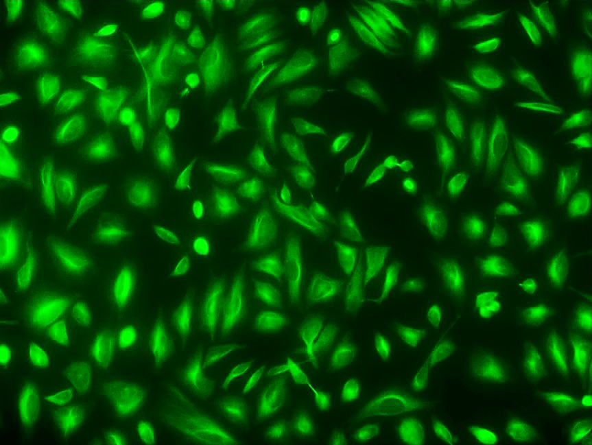
Cell Navigator® Live Cell Tubulin Staining Kit (Green Fluorescence) provides a high-performance solution for live-cell microtubule visualization using Tubulite™ Green, a cell-permeable fluorescent probe designed for imaging tubulin structures. Once inside the cell, Tubulite™ Green is activated by intracellular esterases, transforming into a charged, membrane-impermeant form that selectively binds to tubulin and remains retained for long-term imaging.
Emitting in the green fluorescence channel, this tubulin staining kit is ideal for multiplex imaging with other fluorescent stains (emitting in red or blue regions) and nuclear stains like DAPI. It is optimized for live-cell cytoskeletal imaging, allowing researchers to study tubulin polymerization, cytoskeletal rearrangement, and drug-induced microtubule changes under physiological conditions.
Example protocol
AT A GLANCE
- Prepare cells with test compounds at a density of 5 × 10⁵ to 1 × 10⁶ cells/mL.
- Prepare and add Tubulite™ Green working solution to cells.
- Incubate at 37 °C for 30 to 60 minutes.
- Read fluorescence intensity with FITC filter set.
Important Note: Thaw each kit component at room temperature before starting the experiment.
Note: This protocol only provides a guideline, and should be modified according to your specific needs.
Note: Tubulite™ Green does not stain formaldehyde-fixed cells. Cells cannot be fixed after staining with Tubulite™ Green as fixation alters the structure of microtubules.CELL PREPARATION
For guidelines on cell sample preparation, please visit:
https://www.aatbio.com/resources/guides/cell-sample-preparation.html
PREPARATION OF STOCK SOLUTIONS
Unless otherwise noted, all unused stock solutions should be divided into single-use aliquots and stored at -20 °C after preparation. Avoid repeated freeze-thaw cycles
Add 25 µL DMSO (Component D) into the vial of Tubulite™ Green (Component A), and mix well.
Note: Aliquot and store the unused Tubulite™ Green stock solution at -20 °C. Avoid repeated freeze/thaw cycles.
PREPARATION OF WORKING SOLUTION
Add 2.5 µL of Tubulite™ Green stock solution stock solution and 100 µL 25 mM ReadiUse™ probenecid (Component D) into 1 mL of Assay Buffer (Component B) or buffer of your choice, and mix well.
Note: For best results, this solution should be used within a few hours of its preparation.
SAMPLE EXPERIMENTAL PROTOCOL
Prepare cell samples as per need.
Remove the cell growth medium and wash cells with PBS (Not provided) or any other buffer of your choice (Optional).
- Add 100 µL Tubulite™ Green working solution and incubate them at 37 °C incubator for 30 to 60 minutes.
Note: The appropriate incubation time depends on the individual cell type and cell concentration used. Optimize the incubation time for each experiment.
Remove the working solution and wash cells twice with PBS or any other buffer of your choice supplemented with 2.5 mM probenecid (diluted from Component C).
Cover the cells with an Assay Buffer containing 2.5 mM probenecid (prepared by diluting Component C), and then monitor fluorescence intensity using a fluorescence microscope with a FITC filter set.
References
Authors: Park, Jung Min and Choi, Seock Hwan and Lee, Eun-Shil and Gum, Sang-Il and Hong, Sungkuk and Kim, Dong Sun and Han, Man-Hoon and Lee, Soung-Hoon and Oh, Ji Won
Journal: Tissue engineering and regenerative medicine (2024): 1037-1048
Authors: El Jailani, Safia and Wassmann, Katja and Touati, Sandra A
Journal: Methods in molecular biology (Clifton, N.J.) (2024): 211-227
Authors: Jenks, Andrew D and Fivaz, Marc and Tanos, Barbara E
Journal: Bio-protocol (2021): e4248
Authors: Wen, Gang and Vanheusden, Marisa and Acke, Aline and Valli, Donato and Neely, Robert K and Leen, Volker and Hofkens, Johan
Journal: ACS nano (2020): 7860-7867
Authors: Yi, Kyung Sik and Choi, Chi-Hoon and Jung, Cheolkyu and Lee, Youngjeon and Jeon, Chang-Yeop and Yeo, Hyun-Gu and Ahn, Yujin and Hwang, Jinwoo and Lee, Hong Jun and Cho, Janggeun and Kwak, Bitnarae and Kwak, Kyung A and Lee, Sang-Rae and Cha, Sang-Hoon
Journal: Journal of neuroscience methods (2020): 108838