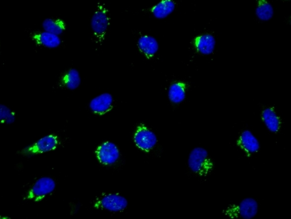
Cell Navigator® NBD Ceramide Golgi Staining Kit *Green Fluorescence*
Price | |
Catalog Number | |
Unit Size | |
Quantity |
Telephone | 1-800-990-8053 |
Fax | 1-800-609-2943 |
sales@aatbio.com | |
International | See distributors |
Bulk request | Inquire |
Custom size | Inquire |
Shipping | Standard overnight for United States, inquire for international |
Excitation (nm) | 467 |
Emission (nm) | 538 |
Certificate of Origin | Download PDF |
H-phrase | H303, H313, H333 |
Hazard symbol | XN |
Intended use | Research Use Only (RUO) |
R-phrase | R20, R21, R22 |
Storage | Freeze (< -15 °C); Minimize light exposure |
UNSPSC | 12352200 |
Overview | ![]() ![]() |
Excitation (nm) 467 | Emission (nm) 538 |
Platform
Fluorescence microscope
Excitation | 488 nm |
Emission | 525 nm |
Recommended plate | Black wall/clear bottom |
Instrument specification(s) | FITC filterset |
Components
Example protocol
AT A GLANCE
- Treat cells as desired
- Add C6 NBD Ceramide working solution and incubate at room temperature or 37°C for 15~30 minutes
- Replace with the Staining Buffer and incubate at room temperature or 37°C for 15~30 minutes
- Observe under microscope using FITC filter set
- Optional: Fix cells with 4% Formaldehyde
Thaw one of each kit component at room temperature before starting the experiment.
CELL PREPARATION
For guidelines on cell sample preparation, please visit https://www.aatbio.com/resources/guides/cell-sample-preparation.html
PREPARATION OF STOCK SOLUTIONS
Unless otherwise noted, all unused stock solutions should be divided into single-use aliquots and stored at -20 °C after preparation. Avoid repeated freeze-thaw cycles
Add 100 µL of DMSO (Component C) into the vial of C6 NBD Ceramide (Component A) and mix well.
Note Store unused C6 NBD Ceramide stock solution at -20o C in single use aliquotes. Avoid freeze-thaw cycles.
PREPARATION OF WORKING SOLUTION
Add 10 µL of NBD Ceramide stock solution (100X) to 990 µL Staining Buffer (Component B) to make NBD Ceramide working solution.
Optional: Add 10 µL Hoechst 33342 (Component D) to 1 mL NBD Ceramide working solution for nuclear stain. Observe under fluorescence microscope with DAPI filter set.
SAMPLE EXPERIMENTAL PROTOCOL
- Plate and treat cells as desired.
Add equal volume of C6 NBD Ceramide working solution directly in cell culture medium. Note: If you have cells in a 96 well plate with 100 µL/well cell culture medium then add 100 µL/well of C6 NBD Ceramide working solution.
- Incubate at room temperature or 37 °C for 15~30min.
- Remove the C6 NBD Ceramide working solution and replace with 100 µL/well of Staining Buffer (Component B).
- Incubate at room temperature or 37 °C for 10~15 minutes.
- Observe under a fluorescence microscope with FITC filter set.
Remove the staining buffer from Step 4, and add 100 µL/well/96-well plate of 4% formaldehyde fixative buffer (not supplied) to each well.
Note For non-adherent cells, add desired amount (such as 2X10⁶ cells/mL) of 4% formaldehyde fixative buffer.
Incubate plates for 20 to 30 minutes at room temperature. Remove fixative. Wash the cells with PBS 2-3 times, and replace with 100 µL/well of Staining Buffer (Component B).
Note The Cell Navigator™ NBD Ceramide Golgi Staining Kit also works well for aldehyde-fixed cells. After fixation, follow the staining protocol steps 2 to 6
Product Family
Name | Excitation (nm) | Emission (nm) |
Cell Navigator® TMR Ceramide Golgi Staining Kit *Red Fluorescence* | 544 | 570 |
Images

Citations
Authors: Venditti, R., Rega, L. R., Masone, M. C., Santoro, M., Polishchuk, E., Sarnataro, D., Paladino, S., D'Auria, S., Varriale, A., Olkkonen, V. M., Di Tullio, G., Polishchuk, R., De Matteis, M. A.
Journal: J Cell Biol (2019): 1055-1065
Authors: Sakamoto, W., Coant, N., Canals, D., Obeid, L. M., Hannun, Y. A.
Journal: J Lipid Res (2018): 2116-2125
Authors: Xie, Z., Hur, S. K., Zhao, L., Abrams, C. S., Bankaitis, V. A.
Journal: Dev Cell (2018): 725-740 e4
Authors: Mitchell, S. B., Iwabuchi, S., Kawano, H., Yuen, T. M. T., Koh, J. Y., Ho, K. W. D., Harata, N. C.
Journal: PLoS One (2018): e0206123
Authors: Ishibashi, Y., Ito, M., Hirabayashi, Y.
Journal: Biochem Biophys Res Commun (2018): 1011-1018
Authors: Rodriguez, M. A., Martinez-Higuera, A., Valle-Solis, M. I., Hern and es-Alej, undefined and ro, M., Chavez-Munguia, B., Figueroa-Gutierrez, A. H., Salas-Casas, A.
Journal: Parasitol Res (2018): 3381-3389
Authors: Liang, Kaifan and Wang, Lirong and Su, Yuqing and Liu, Mengyang and Feng, Rui and Song, Yanzhi and Deng, Yihui
Journal: European Journal of Pharmaceutical Sciences (2018): 210--216
Authors: Back, M. J., Ha, H. C., Fu, Z., Choi, J. M., Piao, Y., Won, J. H., Jang, J. M., Shin, I. C., Kim, D. K.
Journal: Cell Death Dis (2018): 670
Authors: Liu, L. K., Choudhary, V., Toulmay, A., Prinz, W. A.
Journal: J Cell Biol (2017): 131-147
Authors: Capasso, S., Sticco, L., Rizzo, R., Pirozzi, M., Russo, D., Dathan, N. A., Campelo, F., van Galen, J., Holtta-Vuori, M., Turacchio, G., Hausser, A., Malhotra, V., Riezman, I., Riezman, H., Ikonen, E., Luberto, C., Parashuraman, S., Luini, A., D'Angelo, G.
Journal: EMBO J (2017): 1736-1754
Application notes
FAQ
What are the similarities between plant cells and animal cells?
Are Cell Navigator® Cell Plasma Membrane Staining Kits suitable for cell culture medium samples?
Are there any alternatives for ethidium bromide in agarose gels?
Are there any alternatives to Cy5?