logo
AAT Bioquest

Cy7 Styramide *Superior Replacement for Cy7 tyramide*

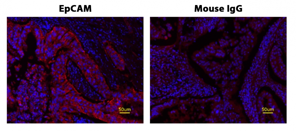
Fluorescence IHC of formaldehyde-fixed, paraffin-embedded human lung adenocarcinoma positive tissue using PSA<strong>&nbsp;&trade;</strong>&nbsp;amplified methods. Human lung adenocarcinoma positive tissue sections were stained with&nbsp;Mouse anti-EpCAM or Control Mouse IgG antibody and then incubated with polyHRP-labeled Goat anti-Mouse IgG secondary antibody followed by Cy7 Styramide&trade; (Cat#45066).
Ordering information
Price
Catalog Number
Unit Size
Quantity
Add to cart
Additional ordering information
Telephone1-800-990-8053
Fax1-800-609-2943
Emailsales@aatbio.com
InternationalSee distributors
Bulk requestInquire
Custom sizeInquire
ShippingStandard overnight for United States, inquire for international
Request quotation
Physical properties
Molecular weight1062.40
SolventDMSO
Spectral properties
Correction Factor (260 nm)0.05
Correction Factor (280 nm)0.036
Correction Factor (482 nm)0.0005
Correction Factor (565 nm)0.0193
Correction Factor (650 nm)0.165
Extinction coefficient (cm -1 M -1)250000
Excitation (nm)756
Emission (nm)779
Quantum yield0.3
Storage, safety and handling
H-phraseH303, H313, H333
Hazard symbolXN
Intended useResearch Use Only (RUO)
R-phraseR20, R21, R22
StorageFreeze (< -15 °C); Minimize light exposure
UNSPSC12171501

OverviewpdfSDSpdfProtocol


See also: Cyanines
Molecular weight
1062.40
Correction Factor (260 nm)
0.05
Correction Factor (280 nm)
0.036
Correction Factor (482 nm)
0.0005
Correction Factor (565 nm)
0.0193
Correction Factor (650 nm)
0.165
Extinction coefficient (cm -1 M -1)
250000
Excitation (nm)
756
Emission (nm)
779
Quantum yield
0.3
Power Styramide™ Signal Amplification (PSA™) system is one of the most sensitive methods that can detect extremely low-abundance targets in cells and tissues with improved fluorescence signal 10-50 times higher than the widely used tyramide (TSA) reagents. In combination with our superior iFluor® dyes that have higher florescence intensity, increased photostability and enhanced water solubility, the iFluor® dye-labeled Styramide™ conjugates can generate fluorescence signal with significantly higher precision and sensitivity (more than 100 times) than standard ICC/IF/IHC. PSA utilizes the catalytic activity of horseradish peroxidase (HRP) for covalent deposition of fluorophores in situ. PSA radicals have much higher reactivity than tyramide radicals, making the PSA system much faster, more robust and sensitive than the traditional TSA reagents. Compared to tyramide reagents, the Styramide™ conjugates have ability to label the target at higher efficiency and thus generate significantly higher fluorescence signal. Styramide™ conjugates also allow significantly less consumption of primary antibody compared to standard directly conjugate method or tyramide amplification with the same level of sensitivity. Cy7 Styramide is optimized to a superior replacement for Cy7 tyramide (Cy7 TSA) or Alexa Fluor® 750 tyramide or other spectrally similar fluorescent tyramide conjugates or TSA reagents.
Platform


Fluorescence microscope

ExcitationCy7 filter set
EmissionCy7 filter set
Recommended plateBlack wall/clear bottom

Example protocol


AT A GLANCE

Protocol Summary
  1. Fix/permeabilize/block cells or tissue
  2. Add primary antibody in blocking buffer
  3. Add HRP-conjugated secondary antibody
  4. Prepare Styramide™ working solution and apply in cells or tissue for 5-10 minutes at room temperature 

PREPARATION OF STOCK SOLUTIONS

Unless otherwise noted, all unused stock solutions should be divided into single-use aliquots and stored at -20 °C after preparation. Avoid repeated freeze-thaw cycles.

1. Cy7 Styramide stock solution (100X)
Add 100 µL of DMSO into the vial of Cy7 Styramide conjugate to make 100X Styramide stock solution.
Note     Make single use aliquots, and store unused 100X stock solution at 2-8 °C in dark place and avoid repeat freeze-thaw cycles.


2. H2O2 stock solution
Add 10 µL of 3% hydrogen peroxide (Not provided) to 90 µL of ddH2O.
Note     Prepare the 100X H2O2 solution fresh on the day of use.

PREPARATION OF WORKING SOLUTION

1. Cy7 Styramide working solution (1X)
Every 1 mL of Reaction Buffer requires 10 µL of Styramide™ stock solution and 10 µL of H2O2 stock solution.
Note     The Styramide provided is enough for 100 tests based on 100 µL of Styramide working solution needed per coverslip or per well in a 96-well microplate.
Note     The Styramide working solution must be used within 2 hours after preparation and avoid direct exposure to light.


2. Secondary antibody-HRP working solution
Make appropriate concentration of secondary antibody-HRP working solution as per the manufacturer's recommendations.

SAMPLE EXPERIMENTAL PROTOCOL

This protocol is applicable for both cells and tissues staining.

Cell fixation and permeabilization
  1. Fix the cells or tissue with 3.7% formaldehyde or paraformaldehyde, in PBS at room temperature for 20 minutes.
  2. Rinse the cells or tissue with PBS twice.
  3. Permeabilize the cells with 0.1% Triton X-100 solution for 1-5 minutes at room temperature.
  4. Rinse the cells or tissue with PBS twice. 

Tissue fixation, deparaffinization and rehydration
Deparaffinize and dehydrate the tissue according to the standard IHC protocols. Perform antigen retrieval with preferred specific solution/protocol as needed.
Protocol can be found at
https://www.aatbio.com/resources/guides/paraffin-embedded-tissue-immunohistochemistry-protocol.html

Peroxidase labeling
  1. Optional: Quench endogenous peroxidase activity by incubating cell or tissue sample in peroxidase quenching solution (such as 3% hydrogen peroxide) for 10 minutes. Rinse with PBS twice at room temperature.
  2. Optional: If using HRP-conjugated streptavidin, it is advisable to block endogenous biotins by biotin blocking buffer.
  3. Block with preferred blocking solution (such as PBS with 1% BSA) for 30 minutes at 4 °C.
  4. Remove blocking solution and add primary antibody diluted in recommended antibody diluent for 60 minutes at room temperature or overnight at 4 °C.
  5. Wash with PBS three times for 5 minutes each.
  6. Apply 100 µL of secondary antibody-HRP working solution to each sample and incubate for 60 minutes at room temperature.
    Note     Incubation time and concentration can be varied depending on the signal intensity.
  7. Wash with PBS three times for 5 minutes each. 

Styramide labeling
  1. Prepare and apply 100 µL of Styramide working solution to each sample and incubate for 5-10 minutes at room temperature.
    Note     If you observe non-specific signal, you can shorten the incubation time with Styramide. You should optimize the incubation period using positive and negative control samples at various incubation time points. Or you can use lower concentration of Styramide in the working solution.
  2. Rinse with PBS three times. 

Counterstain and fluorescence imaging
  1. Counterstain the cell or tissue samples as needed. AAT provides a series of nucleus counterstain reagents as listed in Table 1. Follow the instruction provided with the reagents.
  2. Mount the coverslip using a mounting medium with anti-fading properties.
  3. Use the appropriate filter set to visualize the signal from the Styramide labeling. 
Table 1.Products recommended for nucleus counterstain.
Cat# Product Name Ex/Em (nm)
17548 Nuclear Blue™ DCS1 350/461
17550 Nuclear Green™ DCS1 503/526
17551 Nuclear Orange™ DCS1 528/576
17552 Nuclear Red™ DCS1 642/660

Calculators


Common stock solution preparation

Table 1. Volume of DMSO needed to reconstitute specific mass of Cy7 Styramide *Superior Replacement for Cy7 tyramide* to given concentration. Note that volume is only for preparing stock solution. Refer to sample experimental protocol for appropriate experimental/physiological buffers.

0.1 mg0.5 mg1 mg5 mg10 mg
1 mM94.127 µL470.633 µL941.265 µL4.706 mL9.413 mL
5 mM18.825 µL94.127 µL188.253 µL941.265 µL1.883 mL
10 mM9.413 µL47.063 µL94.127 µL470.633 µL941.265 µL

Molarity calculator

Enter any two values (mass, volume, concentration) to calculate the third.

Mass (Calculate)Molecular weightVolume (Calculate)Concentration (Calculate)Moles
/=x=

Spectrum


Open in Advanced Spectrum Viewer
spectrum

Spectral properties

Correction Factor (260 nm)0.05
Correction Factor (280 nm)0.036
Correction Factor (482 nm)0.0005
Correction Factor (565 nm)0.0193
Correction Factor (650 nm)0.165
Extinction coefficient (cm -1 M -1)250000
Excitation (nm)756
Emission (nm)779
Quantum yield0.3

Product Family


NameExcitation (nm)Emission (nm)Extinction coefficient (cm -1 M -1)
Cy7 tyramide756779250000
Cy7 tetrazine756779250000
PerCP-Cy7757783350000

Images


References


View all 50 references: Citation Explorer
Somatostatin receptor profile in pituitary thyrotroph adenomas.
Authors: Thodou, Eleni and Kontogeorgos, George
Journal: Clinical neurology and neurosurgery (2020): 105865
Identification of Murine Basophils by Flow Cytometry and Histology.
Authors: Schwartz, Christian and Voehringer, David
Journal: Methods in molecular biology (Clifton, N.J.) (2020): 367-375
Use of TAI-FISH to visualize neural ensembles activated by multiple stimuli.
Authors: Zhang, Qi and He, Qiye and Wang, Jihua and Fu, Chaoying and Hu, Hailan
Journal: Nature protocols (2018): 118-133
Selective Inhibition of Amygdala Neuronal Ensembles Encoding Nicotine-Associated Memories Inhibits Nicotine Preference and Relapse.
Authors: Xue, Yan-Xue and Chen, Ya-Yun and Zhang, Li-Bo and Zhang, Li-Qun and Huang, Geng-Di and Sun, Shi-Chao and Deng, Jia-Hui and Luo, Yi-Xiao and Bao, Yan-Ping and Wu, Ping and Han, Ying and Hope, Bruce T and Shaham, Yavin and Shi, Jie and Lu, Lin
Journal: Biological psychiatry (2017): 781-793
Detection of tissue factor-positive extracellular vesicles by laser scanning confocal microscopy.
Authors: Hisada, Yohei and Auriemma, Alyson C and Alexander, Wyeth and Ay, Cihan and Mackman, Nigel
Journal: Thrombosis research (2017): 65-72
Immunohistochemical Methods for Measuring Tissue Lymphangiogenesis.
Authors: Royston, Daniel J and Clasper, Steven and Jackson, David G
Journal: Methods in molecular biology (Clifton, N.J.) (2016): 35-48
Development of a highly sensitive immunohistochemical method to detect neurochemical molecules in formalin-fixed and paraffin-embedded tissues from autopsied human brains.
Authors: Goto, Satoshi and Morigaki, Ryoma and Okita, Shinya and Nagahiro, Shinji and Kaji, Ryuji
Journal: Frontiers in neuroanatomy (2015): 22
TRPM8 has a key role in experimental colitis-induced visceral hyperalgesia in mice.
Authors: Hosoya, T and Matsumoto, K and Tashima, K and Nakamura, H and Fujino, H and Murayama, T and Horie, S
Journal: Neurogastroenterology and motility : the official journal of the European Gastrointestinal Motility Society (2014): 1112-21
Visualizing an emotional valence map in the limbic forebrain by TAI-FISH.
Authors: Xiu, Jianbo and Zhang, Qi and Zhou, Tao and Zhou, Ting-ting and Chen, Yang and Hu, Hailan
Journal: Nature neuroscience (2014): 1552-9
Expression of E6, p53 and p21 proteins and physical state of HPV16 in cervical cytologies with and without low grade lesions.
Authors: Tagle, Diana K Jiménez and Sotelo, Daniel Hernández and Illades-Aguiar, Berenice and Leyva-Vazquez, Marco A and Alfaro, Eugenia Flores and Coronel, Yaneth Castro and Hernández, Oscar Del Moral and Romero, Luz Del Carmen Alarcón
Journal: International journal of clinical and experimental medicine (2014): 186-93