logo
AAT Bioquest

Live or Dead™ Cell Viability Assay Kit *Red/Blue Dual Fluorescence*

Fluorescence images of HeLa cells labeled with Live or Dead™ Cell Viability Assay Kit *Dual Fluorescence* (Cat#22788). HeLa cells at 100,000 cells/well/100 µL were seeded overnight in a 96-well black wall/clear bottom plate. Cells were treated with 0-1 µM staurosporine (SS) at 37ºC for 4 hours (A-D), or fixed in ethanol (E), then incubated with dye-loading solution for 1 hour. The fluorescence signal was measured using a fluorescence microscope with Texas Red or Cy5 filter for viable cells (Red) and DAPI filter for necrotic cells (Blue), respectively. (F) The corresponding fluorescence signal were measured using a FlexStation® microplate reader (Molecular Devices) with bottom read mode at Ex/Em= 610/650 (cutoff=630 nm, Red) and Ex/Em=360/450 (cutoff=420 nm, Blue), respectively.
Ordering information
Price
Catalog Number
Unit Size
Quantity
Add to cart
Additional ordering information
Telephone1-800-990-8053
Fax1-800-609-2943
Emailsales@aatbio.com
InternationalSee distributors
Bulk requestInquire
Custom sizeInquire
ShippingStandard overnight for United States, inquire for international
Request quotation
Spectral properties
Excitation (nm)612
Emission (nm)630
Storage, safety and handling
H-phraseH303, H313, H333
Hazard symbolXN
Intended useResearch Use Only (RUO)
R-phraseR20, R21, R22
UNSPSC12352200

OverviewpdfSDSpdfProtocol


Excitation (nm)
612
Emission (nm)
630
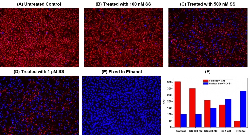
This Live or Dead™ Cell Viability Assay Kit uses two fluorescent indicators: Cellbrite™ Red (Ex/Em = 613/631 nm) for labeling viable cells and a cell-impermeable DNA-binding dye Nuclear Blue™ DCS1 (Ex/Em = 360/450 nm) for labeling dead cells with damaged membranes. Cells grown in black-wall plates can be stained and quantified in less than two hours. The assay is more robust and accurate than the other viability assays. It can be readily adapted for a wide variety of fluorescence platforms such as microplate assays, fluorescence microscopes, and flow cytometry. The kit provides all the essential components with an optimized assay protocol. It is suitable for both proliferating and non-proliferating cells (either suspension or adherent cells).

Platform


Fluorescence microscope

ExcitationCy5 filter (alive), DAPI filter (dead)
EmissionCy5 filter (alive), DAPI filter (dead)
Recommended plateBlack wall/clear bottom

Fluorescence microplate reader

Excitation610, 360 nm
Emission650, 450 nm
Cutoff630, 420 nm
Recommended plateBlack wall/clear bottom
Instrument specification(s)Bottom read mode

Components


Example protocol


AT A GLANCE

Protocol Summary
  1. Prepare cells with test compounds
  2. Add dye-working solution
  3. Incubate at room temperature or 37°C for 30 minutes to 1 hour
  4. Monitor fluorescence intensity (bottom read mode) at Ex/Em = 610/650 nm (Cutoff = 630 nm, Red) and Ex/Em = 360/450 nm (Cutoff =420 nm, Blue) or fluorescence microscope with Cy5 channel (live) and DAPI channel (dead)
Important Note

Thaw all the kit components at room temperature before starting the experiment.

CELL PREPARATION

For guidelines on cell sample preparation, please visit:

https://www.aatbio.com/resources/guides/cell-sample-preparation.html

PREPARATION OF WORKING SOLUTION

Add 5 µL of 200X Cellbrite™ Red (Component A) and 5 µL of 200X Nuclear Blue™ DCS1 (Component C) into 1 mL of Assay Buffer (Component B) and mix well to make dye-working solution. This dye-working solution is stable for at least 1 hour at room temperature.

Note: As the optimal staining conditions may vary depending on different cell types, it’s recommended to determine the appropriate concentration of Component A and C individually.

SAMPLE EXPERIMENTAL PROTOCOL

  1. Prepare cells according to the standard protocol. Note: We treated HeLa cells with staurosporine (SS) for 4 hours at 37ºC to induce cell apoptosis. See Figure 1 for details.
  2. Replace growth medium with 100 µL/well (96-well plate) or 25 µL/well (384-well plate) of dye-working solution.
  3. Incubate the dye-working solution plate at room temperature or 37°C for 30 minutes to 1 hour, protected from light.
  4. Wash cells with HHBS, PBS or buffer of your choice twice.
  5. Add 100 µL/well (96-well plate) or 25 µL/well (384-well plate) of Assay Buffer (Component B) into the cells.
  6. Monitor the fluorescence signal under a fluorescence microscope with Texas Red or Cy5 filter for live cells, and DAPI filter for dead cells. The fluorescence intensity can also be analyzed with a fluorescence microplate reader (bottom read mode) at Ex/Em = 610/650 nm (Cutoff = 630 nm, Red) and Ex/Em = 360/450 nm (Cutoff =420 nm, Blue).

Spectrum


Open in Advanced Spectrum Viewer
spectrum

Spectral properties

Excitation (nm)612
Emission (nm)630

Images


Citations


View all 20 citations: Citation Explorer
Developing a Novel Platelet-Rich Plasma-Laden Bioadhesive Hydrogel Contact Lens for the Treatment of Ocular Surface Chemical Injuries
Authors: Soykan, Merve Nur and Altug, Burcugul and Bas, Harun and Ghorbanpoor, Hamed and Avci, Huseyin and Eroglu, Sertac and Sengel, Sultan Butun and Sariboyaci, Ayla Eker and Bagis, Sibel Gunes and Uysal, Onur and others,
Journal: Macromolecular Bioscience (2023): 2300204
A Multifunctional Sateen Woven Dressings for Treatment of Skin Injuries
Authors: Ozel, Ceren and Apaydin, Elif and Sariboyaci, Ayla Eker and Tamayol, Ali and Avci, Huseyin
Journal: Colloids and Surfaces B: Biointerfaces (2023): 113197
Ultrasound and microbubbles (USMB) potentiated doxorubicin penetration and distribution in 3D breast tumour spheroids
Authors: Misra, Rahul and Rajic, Mathew and Sathiyamoorthy, Krishnan and Karshafian, Raffi
Journal: Journal of Drug Delivery Science and Technology (2020): 102261
Functional imaging of neuronal activity of auditory cortex by using Cal-520 in anesthetized and awake mice
Authors: Li, Jingcheng and Zhang, Jianxiong and Wang, Meng and Pan, Junxia and Chen, Xiaowei and Liao, Xiang
Journal: Biomedical Optics Express (2017): 2599--2610
NINJ2--A novel regulator of endothelial inflammation and activation
Authors: Wang, Jingjing and Fa, Jingjing and Wang, Pengyun and Jia, Xinzhen and Peng, Huixin and Chen, Jing and Wang, Yifan and Wang, Chenhui and Chen, Qiuyun and Tu, Xin and others, undefined
Journal: Cellular Signalling (2017)
Influence of hypothermia and subsequent rewarming upon leukocyte-endothelial interactions and expression of Junctional-Adhesion-Molecules A and B
Authors: Bogert, Nicolai V and Werner, Isabella and Kornberger, Angela and Meybohm, Patrick and Moritz, Anton and Keller, Till and Stock, Ulrich A and Beiras-Fern, undefined and ez, Andres
Journal: Scientific reports (2016)
Inhibition of ABC transport proteins by oil sands process affected water
Authors: Alharbi, Hattan A and Saunders, David MV and Al-Mousa, Ahmed and Alcorn, Jane and Pereira, Alberto S and Martin, Jonathan W and Giesy, John P and Wiseman, Steve B
Journal: Aquatic Toxicology (2016): 81--88
Rapid generation of collagen-based microtissues to study cell--matrix interactions
Authors: Brett, Marie-Elena and Crampton, Alex and ra L , undefined and Wood, David K
Journal: Technology (2016): 1--8
Toxicokinetics and toxicodynamics of chlorpyrifos is altered in embryos of Japanese medaka exposed to oil sands process-affected water: evidence for inhibition of P-glycoprotein
Authors: Alharbi, Hattan A and Alcorn, Jane and Al-Mousa, Ahmed and Giesy, John P and Wiseman, Steve B
Journal: Journal of Applied Toxicology (2016)
Flexible Endoscopic Spray Application of Respiratory Epithelial Cells as Platform Technology to Apply Cells in Tubular Organs
Authors: Thiebes, Anja Lena and Reddemann, Manuel Armin and Palmer, Johannes and Kneer, Reinhold and Jockenhoevel, Stefan and Cornelissen, Christian Gabriel
Journal: Tissue Engineering Part C: Methods (2016): 322--331

References


View all 84 references: Citation Explorer
Functional evidence that the self-renewal gene NANOG regulates esophageal squamous cancer development
Authors: Li, Deng and Xiang, Xiaocong and Yang, Fei and Xiao, Dongqin and Liu, Kang and Chen, Zhu and Zhang, Ruolan and Feng, Gang
Journal: Biochemical and Biophysical Research Communications (2017)
Localized functional chemical stimulation of TE 671 cells cultured on nanoporous membrane by calcein and acetylcholine
Authors: Zibek S, Stett A, Koltay P, Hu M, Zengerle R, Nisch W, Stelzle M.
Journal: Biophys J. (2006)
A vaccination and challenge model using calcein marked fish
Authors: Klesius PH, Evans JJ, Shoemaker CA, Pasnik DJ.
Journal: Fish Shellfish Immunol (2006): 20
Novel fluorescence assay using calcein-AM for the determination of human erythrocyte viability and aging
Authors: Bratosin D, Mitrofan L, Palii C, Estaquier J, Montreuil J.
Journal: Cytometry A (2005): 78
Cytotoxic effects of 100 reference compounds on Hep G2 and HeLa cells and of 60 compounds on ECC-1 and CHO cells. I mechanistic assays on ROS, glutathione depletion and calcein uptake
Authors: Schoonen WG, Westerink WM, de Roos JA, Debiton E.
Journal: Toxicol In Vitro (2005): 505
Calcein AM release-based cytotoxic cell assay for fish leucocytes
Authors: Iwanowicz LR, Densmore CL, Ottinger CA.
Journal: Fish Shellfish Immunol (2004): 127
Calcein-AM is a detector of intracellular oxidative activity
Authors: Uggeri J, Gatti R, Belletti S, Sc and roglio R, Corradini R, Rotoli BM, Orl and ini G., undefined
Journal: Histochem Cell Biol (2004): 499
Comparison of the usefulness of the MTT, ATP, and calcein assays to predict the potency of cytotoxic agents in various human cancer cell lines
Authors: Mueller H, Kassack MU, Wiese M.
Journal: J Biomol Screen (2004): 506
In vitro assay of mineralized-tissue formation on titanium using fluorescent staining with calcein blue
Authors: Goto T, Kajiwara H, Yoshinari M, Fukuhara E, Kobayashi S, Tanaka T.
Journal: Biomaterials (2003): 3885
The effects of calcium chloride and sodium chloride on the electroporation-mediated skin permeation of fluorescein isothiocyanate (FITC)-dextrans in vitro
Authors: Tokudome Y, Sugibayashi K.
Journal: Biol Pharm Bull (2003): 1508