Products
Services
Resources
Selection Guides
About
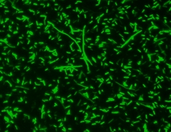
MycoLight™ Green JJ98
5 mM in DMSO
MycoLight™ Green JJ98 is a green-fluorescent nuclear and chromosome stain that is permeant to both prokaryotic and eukaryotic cell membranes. MycoLight™ Green JJ98 has a high affinity for DNA, and exhibits enhanced fluorescence upon binding with an excitation maximum close to the 488 nm argon laser line and fluorescence emission maximum at ~500 nm. MycoLight™ Green JJ98 is particularly useful as a nuclear counterstain for bacterial assays since it stains both live and dead gram-positive and gram-negative bacteria. It is an excellent replacement for SYTO® 9 (SYTO® is the trademark of Invitrogen).
| Catalog | Size | Price | Quantity |
|---|---|---|---|
| 24000 | 100 uL | Price |

Physical properties
| Molecular weight | 534 |
| Solvent | DMSO |
Spectral properties
| Excitation (nm) | 482 |
| Emission (nm) | 512 |
Usage and storage
| Intended use | Research Use Only (RUO) |
| Storage | Freeze (< -15 °C); Minimize light exposure |
Instrument settings
| Flow cytometer | |
| Excitation | 488 nm laser |
| Emission | 530/30 nm filter |
| Instrument specification(s) | FITC channel |
| Fluorescence microscope | |
| Excitation | FITC filter set |
| Emission | FITC filter set |
| Recommended plate | Black wall/clear bottom |
Contact us
| Telephone | |
| Fax | |
| sales@aatbio.com | |
| International | See distributors |
| Bulk request | Inquire |
| Custom size | Inquire |
| Technical Support | Contact us |
| Request quotation | Request |
| Purchase order | Send to sales@aatbio.com |
| Shipping | Standard overnight for United States, inquire for international |
Page updated on July 16, 2026