logo
AAT Bioquest

ReadiUse™ PE [R-Phycoerythrin] *Ammonium Sulfate-Free*

R-Phycoerythrin (PE) is isolated from red algae. Its primary absorption peak is at 565 nm with secondary peaks at 496 and 545 nm. All the commercial PE materials are sold in concentrated ammonium sulfate buffers. The commercial PE materials from other vendors require the tedious dialysis or other purifications performed before it can be used for labeling purposes. AAT Bioquest offers this ReadiUse™ PE that can be readily used for any labelings without any purifications required. Our highly purified ReadiUse™ PE facilitates the rapid PE conjugations to antibodies and other proteins such as streptavidin and other secondary reagents.
Spectrum

Citations

View all 1 citations: Citation Explorer
Targeting pro-inflammatory T cells as a novel therapeutic approach to potentially resolve atherosclerosis in humans
Authors: Fan, Lin and Liu, Junwei and Hu, Wei and Chen, Zexin and Lan, Jie and Zhang, Tongtong and Zhang, Yang and Wu, Xianpeng and Zhong, Zhiwei and Zhang, Danyang and others,
Journal: Cell Research (2024): 1--21

References

View all 46 references: Citation Explorer
Chromophore attachment to phycobiliprotein beta-subunits: phycocyanobilin:cysteine-beta84 phycobiliprotein lyase activity of CpeS-like protein from Anabaena Sp. PCC7120
Authors: Zhao KH, Su P, Li J, Tu JM, Zhou M, Bubenzer C, Scheer H.
Journal: J Biol Chem (2006): 8573
Excitation energy transfer from phycobiliprotein to chlorophyll d in intact cells of Acaryochloris marina studied by time- and wavelength-resolved fluorescence spectroscopy
Authors: Petrasek Z, Schmitt FJ, Theiss C, Huyer J, Chen M, Larkum A, Eichler HJ, Kemnitz K, Eckert HJ.
Journal: Photochem Photobiol Sci (2005): 1016
Single-molecule spectroscopy selectively probes donor and acceptor chromophores in the phycobiliprotein allophycocyanin
Authors: Loos D, Cotlet M, De Schryver F, Habuchi S, Hofkens J.
Journal: Biophys J (2004): 2598
Isolation and characterisation of phycobiliprotein rich mutant of cyanobacterium Synechocystis sp
Authors: Prasanna R, Dhar DW, Dominic TK, Tiwari ON, Singh PK.
Journal: Acta Biol Hung (2003): 113
Evaluation of Tolypothrix germplasm for phycobiliprotein content
Authors: Prasanna R, Prasanna BM, Mohammadi SA, Singh PK.
Journal: Folia Microbiol (Praha) (2003): 59
Page updated on October 24, 2024

Ordering information

Price
Unit size
1 mg
10 mg
Catalog Number
Quantity
Add to cart

Additional ordering information

Telephone1-800-990-8053
Fax1-800-609-2943
Emailsales@aatbio.com
InternationalSee distributors
Bulk requestInquire
Custom sizeInquire
Technical SupportContact us
Purchase orderSend to sales@aatbio.com
ShippingStandard overnight for United States, inquire for international
Request quotation

Physical properties

Molecular weight

~240000

Solvent

Water

Spectral properties

Correction Factor (280 nm)

0.175

Extinction coefficient (cm -1 M -1)

1960000

Excitation (nm)

565

Emission (nm)

574

Quantum yield

0.82

Storage, safety and handling

H-phraseH303, H313, H333
Hazard symbolXN
Intended useResearch Use Only (RUO)
R-phraseR20, R21, R22

Storage

Refrigerated (2-8 °C); Minimize light exposure
UNSPSC12171501
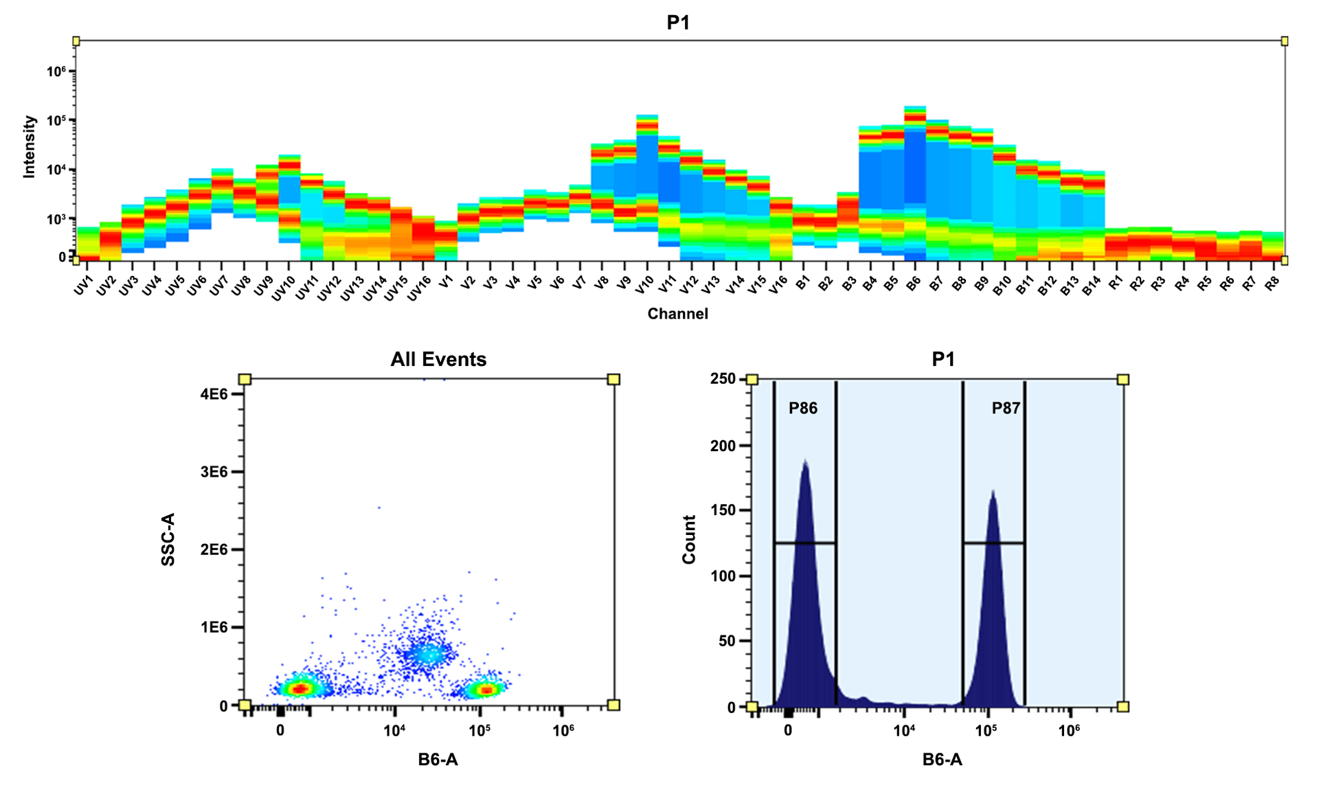
Top) Spectral pattern was generated using a 4-laser spectral cytometer. Spatially offset lasers (355 nm, 405 nm, 488 nm, and 640 nm) were used to create four distinct emission profiles, then, when combined, yielded the overall spectral signature. Bottom) Flow cytometry analysis of whole blood cells stained with PE anti-human CD4 *SK3* conjugate. The fluorescence signal was monitored using an Aurora spectral flow cytometer in the PE specific B6-A channel.
Flow cytometry analysis of whole blood stained with PE anti-human CD4 *SK3* conjugate. The fluorescence signal was monitored using an Aurora spectral flow cytometer in the PE specific B4-A channel.
R-Phycoerythrin (PE) is isolated from red algae.  Its primary absorption peak is at 565 nm with secondary peaks at 496 and 545 nm. AAT Bioquest offers this ReadiUse™ PE that can be readily used for any labelings without any purifications required.